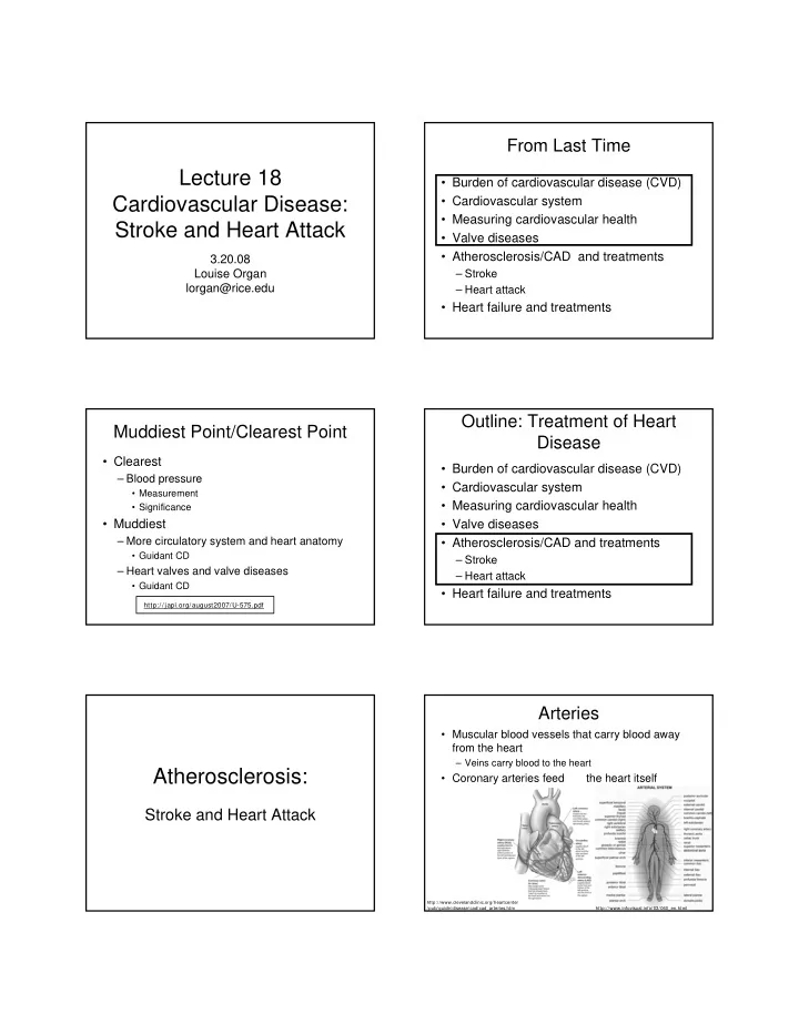
Lecture 18 Burden of cardiovascular disease (CVD) Cardiovascular - - PDF document

lecture 18
SMART_READER_LITE
LIVE PREVIEW

Lecture 18 Burden of cardiovascular disease (CVD) Cardiovascular - - PDF document

From Last Time Lecture 18 Burden of cardiovascular disease (CVD) Cardiovascular Disease: Cardiovascular system Measuring cardiovascular health Stroke and Heart Attack Valve diseases Atherosclerosis/CAD and treatments


slide-1
SLIDE 1

Lecture 18 Cardiovascular Disease: Stroke and Heart Attack

3.20.08 Louise Organ lorgan@rice.edu

From Last Time

  • Burden of cardiovascular disease (CVD)
  • Cardiovascular system
  • Measuring cardiovascular health
  • Valve diseases
  • Atherosclerosis/CAD and treatments

– Stroke – Heart attack

  • Heart failure and treatments

Muddiest Point/Clearest Point

  • Clearest

– Blood pressure

  • Measurement
  • Significance
  • Muddiest

– More circulatory system and heart anatomy

  • Guidant CD

– Heart valves and valve diseases

  • Guidant CD

http://japi.org/august2007/U-575.pdf

Outline: Treatment of Heart Disease

  • Burden of cardiovascular disease (CVD)
  • Cardiovascular system
  • Measuring cardiovascular health
  • Valve diseases
  • Atherosclerosis/CAD and treatments

– Stroke – Heart attack

  • Heart failure and treatments

Atherosclerosis:

Stroke and Heart Attack

Arteries

  • Muscular blood vessels that carry blood away

from the heart

– Veins carry blood to the heart

  • Coronary arteries feed

the heart itself

http://www.clevelandclinic.org/heartcenter /pub/guide/disease/cad/cad_arteries.htm http://www.infovisual.info/03/060_en.html

slide-2
SLIDE 2

Artery Anatomy Atherosclerotic Plaque

http://www.nhlbi.nih.gov/health/dci/Diseases/Hbc/HBC_WhatIs.html http://medlib.med.utah.edu/WebPath/jpeg5/CV119.jpg http://www.brown.edu/Courses/Digital_Path/He art/atherosclerosis.htm

Atherosclerosis: Histology Atherosclerosis: Two Problems

  • Occlusion (Stenosis)

– Narrowed artery diameter – Less blood flow

  • Thrombus

– Formation of clots inside blood vessels – Can completely block blood flow

Atherosclerosis: Thrombus

  • The thin layer or “cap” covering a plaque

can rupture or burst

– The inside of the plaque is then exposed to blood – This sends chemical signals to a host of blood cells

  • Platelets aggregate to form a clot

– Thrombus can block blood flow – Not enough oxygen delivered -- ischemia – Tissues can begin to die -- infarction

http://www.medimagery.com/pathology.jpeg

Thrombus Development

http://www.nlm.nih.gov/medlineplus/ency/imagepages/18020.htm

slide-3
SLIDE 3

Atherosclerosis: Embolus

  • Thrombus can be displaced from the origin

and travel to another location

– Vessels shrink in diameter away from the heart

  • Can then lodge in normally healthy vessels

– Can cause blockage in distant location -- embolus

  • If vessels in the neck or head are affected

– Ischemia or infarction of the brain tissue – Stroke

Stroke (Cerebrovascular Event)

  • A leading cause of death in older

Americans

  • # 1 cause of adult disability
  • Caused by thrombus or embolus in

vessels feeding brain

  • Infarction results in brain damage
  • Effects and long term damage depends on

area of infarction

  • Two-thirds of survivors have a disability

– Limb weakness, speech impediment,

http://www.stroke.org/site/PageNavigator/HOME

Stroke: Risk Factors

  • High blood pressure
  • High cholesterol levels
  • Tobacco use
  • Excessive alcohol

consumption

  • Diabetes
  • Sedentary lifestyle
  • Poor diet/nutrition

Stroke: Symptoms

  • Stroke has a very rapid onset
  • Sudden appearance of the following

symptoms warrants medical attention

– Numbness or weakness of face, arm or leg - especially on one side of the body – Confusion, trouble speaking or understanding – Trouble seeing in one or both eyes – Trouble walking, dizziness, loss of balance or coordination – Severe headache with no known cause

Stroke: “Act F.A.S.T”

  • Face

– Ask the person to smile

  • Does one side of the face droop?
  • Arms

– Ask the person to raise both arms

  • Does one arm drift downward?
  • Speech

– Ask the person to repeat a simple sentence

  • Are word slurred? Can they repeat correctly?
  • Time

– Time is important!

  • Call 911 and/or seek medical help immediately

Heart Attack

slide-4
SLIDE 4

Atherosclerosis vs. Coronary Artery Disease (CAD)

  • Atherosclerosis

– The build-up of plaques throughout vessel systems – Can have effects at distant sites

  • CAD

– Plaques in the coronary arteries – Specifically affects the heart muscle

Atherosclerosis CAD Clot stops flow of blood Plaque build up

  • n vessel walls

Where the blockage

  • ccurs in the artery

Part of the heart affected

Coronary Atherosclerotic Plaque Early Warning Signs: Angina

  • Plenty of people with CAD have no

symptoms

  • Most prevalent symptom is angina

– Chest pain

  • Two types of angina: stable and unstable

– Stable is brought on by stress or exercise – Unstable may be sudden or more variable

  • Angina is typically the result of ischemia

– Heart needs more oxygen

Progression of Heart Disease

High Blood Pressure High Cholesterol Levels Atherosclerosis Ischemia Heart Attack Heart Failure

CAD and Heart Attacks

  • Myocardial infarction (MI or AMI)

– Myocardium: heart muscle – Infarction: tissue death due to lack of blood (oxygen)

  • Pathophysiology

– Functional changes associated with or resulting from MI

  • Diagnosis
  • Treatment

Guidant Heart Attack

MI Pathophysiology: Case Study

  • Three months following his first visit to your office, Mr.

Solomon presents to the ER in the early morning, with chest pain of one hour duration.

  • Mr. Solomon describes the pain as being severe and "like

someone was sitting on his chest." The pain, located "in the lower part of my breast bone," awakened him from his sleep. Although he tried to relieve the pain by changing positions in bed, sitting up and drinking water, it remained unchanged.

  • He did not sleep well because "I had an upset stomach an

acid-burning feeling." He attributed these symptoms to over eating and drinking at a Christmas party.

  • He has no pain or discomfort in his arms but says he has an

"achiness" in his left jaw which he attributes to "bad teeth."

  • Physical examination reveals the patient to be anxious, pale,

diaphoretic and in obvious discomfort. He is unshaven and accompanied by his wife. He tries to relieve his pain by

  • belching. He coughs occasionally. Mr. Solomon says "the flu

has been going around the office, and I’ve had a little cough and fever all week.”

slide-5
SLIDE 5

Warning Signs of Heart Attack

  • Many heart attacks start slowly; symptoms may come

and go

  • Chest discomfort

– Most heart attacks involve discomfort in the center of the chest that lasts for more than a few minutes, or goes away and comes

  • back. The discomfort can feel like uncomfortable pressure,

squeezing, fullness, or pain

  • Discomfort in other areas of the upper body

– Can include pain or discomfort in one or both arms, the back, neck, jaw, or stomach

  • Shortness of breath

– Often comes along with chest discomfort. But it also can occur before chest discomfort

  • Other symptoms

– May include breaking out in a cold sweat, nausea, or light- headedness

http://www.archive.org/details/gov.hhs.nih.56-042n

Take Home Points

  • Everyone is different

– Symptoms will vary

  • You don’t have to be “old” to have a heart

attack

  • Women and men are equally susceptible
  • The speed with which you receive

treatment can make the difference

  • Importance of prevention!

US Burden of Heart Attack

  • 1.1 million/year
  • 460,000 of those heart attacks are fatal

– ~ 40%

  • Half of those deaths occur within 1 hour of

symptom onset, before person reaches hospital

– High mortality highlights the importance of prevention – And diagnosis if you have angina or other symptoms

Heart Attack: Diagnosis of Atherosclerosis

  • Blood tests
  • Electrocardiograms (ECG)
  • Ultrasound (echocardiogram)
  • Stress tests
  • Coronary angiography
  • Computer tomography (CT)

Atherosclerosis Diagnosis: Angiography

  • Catheter threaded up

to heart

  • Contrast agent is

injected near problem area

– Blood looks same as tissues to X-rays

  • Use standard X-ray or

CT to take pictures and image occlusions

http://www.nlm.nih.gov/medlineplus/ency/ imagepages/18129.htm

Angiography: Hardware

slide-6
SLIDE 6

Angiography: Images

Atherosclerosis and Heart Attacks

Treatment of Acute Occlusion

Treatment of Coronary Occlusions

  • Introduced in Lecture 4
  • Thrombolytic drugs
  • Angioplasty
  • Stents

– Often in combination with angioplasty

  • Coronary artery bypass graft (CABG)
  • Which one would you choose?

Thrombolytic Drugs

  • Tissue plasminogen activator (tPA):

– Protease that dissolves blood clots – Approved for use in certain heart attack or stroke patients

  • Clinical Studies:

– tPA and other clot-dissolving agents can reduce the amount of damage to the heart muscle and save lives – To be effective, they must be given within a few hours after symptoms begin

  • Few MI patients qualify

– Administered through an intravenous (IV) line in the arm by hospital personnel – Patients treated within 90 min after onset of chest pain are 1/7 as likely to die compared to patients who receive therapy later

Thrombolytic Drugs

  • Risks of thrombolytics:

– Intra-cranial hemorrhage

  • Increased risk in those > age 70
  • Instead of ischemic stroke this can lead to

hemorrhagic stroke

– Patients may require further intervention

  • Costs of thrombolytics:

– tPA = $2300 – Streptokinase = $320

Effectiveness of Thrombolytics

  • Clinical Trial:

– In 15 countries and 1081 hospitals – 41,021 patients with evolving myocardial infarction – Randomly assigned to 4 different strategies:

  • Streptokinase and subcutaneous heparin
  • Streptokinase and IV heparin
  • Accelerated tissue plasminogen activator (t-PA) and IV heparin
  • Combo of streptokinase plus t-PA with IV heparin

– Primary end point was 30-day mortality

  • Result:

– Streptokinase & subcutaneous heparin: 7.2% (stroke 0.49%) – Streptokinase & IV heparin: 7.4% (stroke 0.54%) – Accelerated t-PA & IV heparin: 6.3% (stroke 0.72%) – Combo of both with IV heparin: 7 0% (stroke 0 94%)

slide-7
SLIDE 7

Cost-Effectiveness of Thrombolytics

Therapy Patient Group $ per yr life saved

tPA Post MI, high risk $3,600 tPA Acute MI, large infarct, treatment started > 2 hours post $24,200 Counseling Smoking cessation $1,300-$3,900 CABG Two vessel disease, severe angina $9,200-$42,500

http://www.sciencedirect.com/science?_ob= ArticleURL&_aset= B-WA-A-A-A-MsSAYZA-UUA AUYWDCBYZYAUYUBBVZZYBWAUBWEUBAU&_rdoc= 1&_fmt= full&_udi= B6T1048NJXK25&_coverDate= 5% 2F22%2F2003&_cdi= 487 6&_orig= search&_st= 13&_sort= d&view= c&_acct= C000004378&_version 1&_urlVersion= 0&_userid= 108429&md5= 5f493caa5f65762c23c0d90eaea8b92d

Treatment of Atherosclerosis

  • Percutaneous Transluminal Angioplasty

(PCTA)

– Balloon Angioplasty

PCTA: Effectiveness

  • Cannot always successfully perform

procedure

– Diffuse disease – Total occlusion – Calcified disease

  • Hardening of the arterial walls
  • Restenosis

– Occurs in 25-54% of patients – Usually occurs within 6 months

slide-8
SLIDE 8

Treatment of Atherosclerosis: Stents

  • A wire (typically) cage is inserted along

with a balloon

– Incorporates angioplasty

  • Inflate the balloon
  • Place the stent
  • Retract balloon
  • Stent remains

Stents Stents Stents

  • Are stents more effective than

angioplasty?

  • The cage does provide support for the

vessel

  • Restenosis is still a major concern
  • Endothelial cells proliferate and grow over

and around the stent

Drug-Eluting Stents

  • Slowly releases drugs

– Hinder cell proliferation around and over the stent – Slow restenosis – May increase thrombosis

  • FDA approved for new lesions in small vessels
  • More expensive (3-4 times)
  • Very popular with public

– NPR story from October 2003

  • Long-term effectiveness

– Restenosis is pronounced in small vessels

  • The verdict is still out

http://www.npr.org/features/feature.php?wfId= 1452217

Treatment of Atherosclerosis

  • Coronary artery bypass graph (CABG)
  • A graft vessel from another area of the

body is used to circumvent the occlusion and return blood flow

  • To do this we

we have to stop the heart from beating

http://www.nlm.nih.gov/medlin eplus/tutorials/coronaryarteryby passgraft/htm/index.htm

slide-9
SLIDE 9

CABG Procedure

  • 1. Patient is prepped, general anesthesia
  • 2. Chest access is gained, through sternum
  • 3. Graft vessel is retrieved
  • 4. Expose heart through pericardium
  • 5. Divert blood through heart lung machine
  • 6. Stop heart
  • 7. Insert graft
  • 8. Return circulation to heart
  • 9. Close incision

Heart-Lung (Bypass) Machine

  • Connected to patient by a series of tubes

that the surgical team places

  • Consists of a chamber that receives the

blood from the body

  • Blood is pumped by machine through an
  • xygenator
  • Oxygenator removes CO2 and adds
  • xygen
  • Pump then pumps this newly oxygenated

blood back to the body

Heart Lung Machine

slide-10
SLIDE 10

Heart Lung Machine Heart Lung Machine CABG Effectiveness

  • 2001: 516,000 CABG procedures performed
  • Procedure takes 4-6 hours, 5-7 day hospital stay
  • Grafts remain open & functioning for 10-15 yrs

– Little restenosis

  • Risks:

– Heart attack (5%) – Stroke (5%) (risk greatest in those over 70 years old) – Death (1-2%) – Sternal wound infection (1-4%) – “Post-pericardiotomy syndrome“ (30%)

  • Occurs few days to 6 months after surgery
  • Symptoms are fever and chest pain

– Some people report memory loss and loss of mental clarity or "fuzzy thinking" following CABG

Innovations

  • Off-pump CABG:

http://www.surgery.usc.edu/divisions/ct/videos-mpeg-

  • ffpumpcoronaryarterybypassgrafting.html
  • Closed chest CABG:

http://www.hsforum.com/stories/storyReader$1537

Comparison of Treatment Methods

  • Hospital Stay:

– CABG: 4-7 days – PCTA: 1-2 days – Stent: 1-2 days

  • Restenosis:

– CABG: 5-6%, usually after 5 years – PCTA : 25-45%, usually within 6 months – Stent: 15-20%, usually within 6 months

Comparison of RX Methods

  • Cost

– CABG: $35,000 – PCTA : $17,000 – Stent: $19,000

  • With drug-eluting stent: $30,000
  • Cost-effectiveness

– Additive procedures:

  • Within 5 years, 20-40% of patients have second PCTA, 25%

have CABG

– Additive costs:

  • 0 years: per patient costs of PCTA 30-50% those of CABG
  • 1 year: 50-60%
  • 3 years: 60-80%
  • >3 years: >80%
slide-11
SLIDE 11

Prevention versus Treatment?

  • Which is more cost effective in terms of

both money and resources?

– For the patient? – For medical community? – For society?

http://www.nytimes.com/2004/03/21/health/21HEAR.html

Progression of Heart Disease

High Blood Pressure High Cholesterol Levels Atherosclerosis Ischemia Heart Attack Heart Failure

Assignments Due Next Tuesday

  • Project Task 5
  • Muddiest Point/Clearest Point