
SLIDE 1
IMA Workshop - "Novel CT Data Acquisition and Processing" 10/16/2019 Web Stayman, Advanced Imaging Algorithms and Instrumentation Lab (aiai.jhu.edu) 1
- J. Webster Stayman
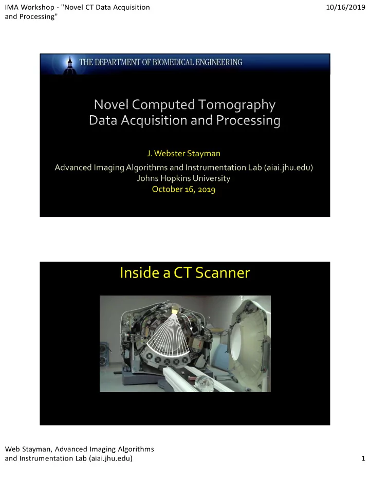
Inside a CT Scanner Web Stayman, Advanced Imaging Algorithms and - - PDF document
IMA Workshop - "Novel CT Data Acquisition 10/16/2019 and Processing" J. Webster Stayman Advanced Imaging Algorithms and Instrumentation Lab (aiai.jhu.edu) Johns Hopkins University October 16, 2019 Inside a CT Scanner Web Stayman,
IMA Workshop - "Novel CT Data Acquisition and Processing" 10/16/2019 Web Stayman, Advanced Imaging Algorithms and Instrumentation Lab (aiai.jhu.edu) 1
IMA Workshop - "Novel CT Data Acquisition and Processing" 10/16/2019 Web Stayman, Advanced Imaging Algorithms and Instrumentation Lab (aiai.jhu.edu) 2
https://www.youtube.com/watch?v=2CWpZKuy-NE
Mean measurements as a function of parameters
IMA Workshop - "Novel CT Data Acquisition and Processing" 10/16/2019 Web Stayman, Advanced Imaging Algorithms and Instrumentation Lab (aiai.jhu.edu) 3
100 200 300 400 500 600 700 100 200 300 400 500 600 700
0.02 0.04 0.06 0.08 100 200 300 400 500 600 700 100 200 300 400 500 600 700 1000 2000 3000 4000 5000 6000 7000 8000 9000 10000
Detector Patient X-ray Source
IMA Workshop - "Novel CT Data Acquisition and Processing" 10/16/2019 Web Stayman, Advanced Imaging Algorithms and Instrumentation Lab (aiai.jhu.edu) 4
Moiré patterns
Mathijs Delbaere, behance.net
(Stayman et al., SPIE 2016)
Detector Patient MAD Filter X-ray Source
X-rays 135 mm 15 mm Thickness: 2mm
MAD0 MAD1
Spacing: 10 mm
Manufacturing:
Tungsten powder, Laser Sintering, EDM Wire cutting
Design Target:
Flatten fluence behind 1) a QRM phantom (30 x 20 cm), 2) cylinders 20~50 cm in diameter
Stayman, SPIE 2016 ; Mathews, CT Meeting 2016, SPIE 2017 ; Gang, PMB 2019
MAD0 MAD1
1 cm X-ray Source MADs Motion Stage Flat Panel Detector 0 cm SMD = 34 cm SAD = 80 cm SDD = 108 cm
Actuation Stages
Motion system on CT gantry
Beam shape: Relative displacement between MADs Amplitude: View-dependent mA and/or ms modulation Miscentered object* and VOI imaging**: Both MADs moving to change beam center
*Mao, SPIE 2018, JMI 2018; ** Wang, CT Meeting 2018, SPIE 2019, JMI 2019
Linear motors
IMA Workshop - "Novel CT Data Acquisition and Processing" 10/16/2019 Web Stayman, Advanced Imaging Algorithms and Instrumentation Lab (aiai.jhu.edu) 5
Uniform Elliptical acrylic phantom (25.8 x 14.1 cm) MAD gain in air Phantom acquisition “One period” of MAD profiles Actuation for elliptical phantom
Prior Information about Patient Initial Ultra-Low Dose Scan Custom Acquisition
Patient-Driven Diagnostic CT 3D Scout Volume Customized Tube Current Modulation Customized Model-based Reconstruction Custom Regularization
r1 r2 r3 r4
𝜈 = argmax Φ 𝑧; 𝜈 Φ = 𝑀 𝑧, 𝜈 − 𝑆(𝜈)
IMA Workshop - "Novel CT Data Acquisition and Processing" 10/16/2019 Web Stayman, Advanced Imaging Algorithms and Instrumentation Lab (aiai.jhu.edu) 6
True Signal Added noise realizations with different correlation while maintaining same variance “noise masquerading as signal” Contrary to CNR, task-based metrics: Accounts for spatial frequency characteristics of task, noise and system response Accounts for observer detection strategy and detection threshold
IMA Workshop - "Novel CT Data Acquisition and Processing" 10/16/2019 Web Stayman, Advanced Imaging Algorithms and Instrumentation Lab (aiai.jhu.edu) 7
Spatial resolution
Detectability Index (non-prewhitening observer)
Noise Imaging task 𝑒
Ω, Ω =
∫ ∫ ∫ 𝑁𝑈𝐺
2 Ω, Ω ⋅ 𝑋2𝑒𝑔 𝑒𝑔 𝑒𝑔
Ω, Ω 𝑁𝑈𝐺 2 Ω, Ω ⋅ 𝑋2𝑒𝑔 𝑒𝑔 𝑒𝑔
𝑦 𝑧
𝑋
𝑔
Hypothesis #1 Hypothesis #2
Hypothesis #1 Hypothesis #2
Quadratic Penalty Likelihood
𝜈 = argmax log 𝑀 𝜈; 𝑧 − 𝛾𝑆 𝜈
Penalized Likelihood Estimation (PLE)
𝑂𝑄𝑇
≈
ℱ AD 𝑧 A𝑓
A𝑓
+ 𝛾𝐒𝑓
(via the projection data)
𝑁𝑈𝐺
≈
ℱ AD 𝑧 A𝑓
A𝑓
+ 𝛾𝐒𝑓
:
Fessler, IEEE-TIP 5(3), (1996); Stayman and Fessler, Trans. Med. Im. 23(12),2004; Zhang-O’Connor and Fessler, IEEE, 2007;
ej : Location Dependence
Forward Model:
𝛾𝐒: Regularization dependence
Gang et al., Med Phys 41(8) 2014
Local NPS and MTF
(1) (2) (3)
𝑧 = 𝐽 𝑣, 𝜄 𝑓
𝑣: Detector location, q: Projection angle
NPS MTF
x10-4
0.5 1.0 1.2
(1) (2) (3)
0.4
fx
0.4
fy
0.4
fx
0.4
fx
0.4
fy
IMA Workshop - "Novel CT Data Acquisition and Processing" 10/16/2019 Web Stayman, Advanced Imaging Algorithms and Instrumentation Lab (aiai.jhu.edu) 8
Task-Driven Optimization System Model Objective Optimizer
Low Dose 3D Scout
Location Contrast Spatial frequency
argmax
,
𝑒 Ω, Ω 𝑁𝑈𝐺 Ω, Ω Spatial resolution: Noise: 𝑂𝑄𝑇 Ω, Ω
Detectability index
Imaging Task
Anatomical Model
𝑒′ Ω, Ω
∗
Ω Ω
mAs, kV, Orbit Fluence field Kernel (FBP) Regularization (MBIR) Conventional Task-driven
0o
90o 180o 270o
0.8 mAs 0.6 0.4 0.2
r1 r2 r3 r4
∗
Task-Driven Imaging
0.65
−0.05 0.37 −1.07
1.12 0.50
Unmodulated Uniform Signal Minimum Variance* (FBP) Task-driven TCM Task-driven Regularization Task-driven TCM + Reg.
90° 0° 180° 270° 0° 180° 0° 180° 0° 180° 0° 180° 0° 180° *Gies et al, MedPhys, 1999
IMA Workshop - "Novel CT Data Acquisition and Processing" 10/16/2019 Web Stayman, Advanced Imaging Algorithms and Instrumentation Lab (aiai.jhu.edu) 9
Unmodulated Uniform Signal Minimum Variance (FBP) Task-driven TCM Task-driven Anisotropic Regularization Task-driven TCM + Aniso Reg.
𝑒′ = 0.77 𝑒
𝑒′ = 0.90 𝑒′ = 1.08 𝑒′ = 1.0 𝑒′ = 1.10
All recons: are quadratic penalized likelihood have the 3 target stimulus have optimal b selection
Phantom Task and Phantom Definition rij(x,y) Task Function Stimulus b (x,y) Flattened Fluence
Task-Driven Design Objective
argmax
,
min 𝑒
Ω, Ω
𝑒
Ω, Ω
⋮ 𝑒
Ω, Ω
IMA Workshop - "Novel CT Data Acquisition and Processing" 10/16/2019 Web Stayman, Advanced Imaging Algorithms and Instrumentation Lab (aiai.jhu.edu) 10
Unmodulated a = 0.5 Task-Driven a = 1.0
Detectability Maps
"Task-driven optimization of fluence field and regularization for model-based iterative reconstruction in computed tomography", IEEE Transactions on Medical Imaging (Special Issue on Low-Dose CT), 36(12), 2424-35 (December 2017)
IMA Workshop - "Novel CT Data Acquisition and Processing" 10/16/2019 Web Stayman, Advanced Imaging Algorithms and Instrumentation Lab (aiai.jhu.edu) 11
Preoperative Image Planning Data Task Definition Conventional Intraoperative CT
X-ray Source Flat-Panel Detector
Traditional Circular Trajectory
Interventional Imaging Conventionally Ignored by Interventional Devices
Task-Driven Trajectory
Prior Information about Patient and Task
Patient- and Task-Driven Intraoperative CT Diagnostic Imaging
IMA Workshop - "Novel CT Data Acquisition and Processing" 10/16/2019 Web Stayman, Advanced Imaging Algorithms and Instrumentation Lab (aiai.jhu.edu) 12
Anthropomorphic Head Phantom and Synthetic Vasculature CBCT Testbench with 6DOF Object Platform
Circular Scan Task-Driven Trajectory Preoperative Scan CBCT Bench Results
IMA Workshop - "Novel CT Data Acquisition and Processing" 10/16/2019 Web Stayman, Advanced Imaging Algorithms and Instrumentation Lab (aiai.jhu.edu) 13
Optimization:
30 stimulus locations on ellipsoid surrounding embolization coil 9 orbital bases
0° ≤ q ≤ 360° CMA-ES (pop=40)
2 2 2 1 2 (1) (2 ( ) , )
arg max min ' ; , ' , , , ; ,..., ' ; ˆ ˆ ˆ ,
Task Task Task L L
d d d W W W a a a a a a a
F
F F F F
IMA Workshop - "Novel CT Data Acquisition and Processing" 10/16/2019 Web Stayman, Advanced Imaging Algorithms and Instrumentation Lab (aiai.jhu.edu) 14
Circular Scan Task-Driven Trajectory
0.014 0.016 0.018 0.02 0.022 0.024 0.026 0.028
Spectral CT involves measurements with varied spectral sensitivity and allows for enhanced material discrimination. Single-shot multi-phasic studies New contrast agent development
Gold Nanoparticles (angiography, lung cancer) Bismuth Sulphide (lymph nodes) Tantalum Oxide (cartilage, lymph nodes) Xenon (lung ventilation)
Symons R, Krauss B, Sahbaee P, Cork TE, Lakshmanan MN, Bluemke DA, Pourmorteza A. Photon-counting CT for simultaneous imaging of multiple contrast agents in the abdomen: An in vivo study. Med Phys. 2017 Oct 26 44(10):5120–5127.
Spectral CT with Photon Counting Detectors
IMA Workshop - "Novel CT Data Acquisition and Processing" 10/16/2019 Web Stayman, Advanced Imaging Algorithms and Instrumentation Lab (aiai.jhu.edu) 15
𝑧 = 𝑇 exp − 𝑟
Φ = 𝑀 𝑧; 𝜍 + 𝑆 𝑀 𝑧; 𝜍 = 𝑥 𝑧 𝜍 − 𝑧
X-ray Focal Spot Spatial-Spectral Filter Multi-Contrast Phantom Energy- Integrating Detector
Overall System Geometry Varied Spectral Beam-lets
Stayman JW and Tilley II S (May 2018) Model-based multi-material decomposition using spatial-spectral filters CT Meeting 2018 102-105. Filtered Beamlets Pb Au Lu Er Polyenergetic Incident Beam
K-Edge Filter Tiles Translating Tiled Filter
Filter Details Data Collection
IMA Workshop - "Novel CT Data Acquisition and Processing" 10/16/2019 Web Stayman, Advanced Imaging Algorithms and Instrumentation Lab (aiai.jhu.edu) 16
I Gd Au Tivnan M, Wang W, Tilley II S, Stayman JW (July 2019) Designing Spatial-Spectral Filters for Spectral CT AAPM Annual Meeting
Sb Er W PbSbErW Pb
IMA Workshop - "Novel CT Data Acquisition and Processing" 10/16/2019 Web Stayman, Advanced Imaging Algorithms and Instrumentation Lab (aiai.jhu.edu) 17
Teflon ~1900mg/ mL (x7) ~1000mg/ mL Solid Water 2mg/mL I 5mg/mL I 7.5mg/mL I 10mg/mL I 15mg/mL I 20mg/mL I Teflon
Spatial-Spectral Filters
Filter Tile Width: 2mm /10 pixels
Customized data acquisitions allow task-based optimization Image quality assessments should take the imaging task into account Understanding and prediction of image properties can drive customization Challenges:
IMA Workshop - "Novel CT Data Acquisition and Processing" 10/16/2019 Web Stayman, Advanced Imaging Algorithms and Instrumentation Lab (aiai.jhu.edu) 18
Collaborators Grace Gang – Biomedical Engineering Kelvin Hong - (Interventional) Radiology Satomi Kawamoto - Radiology A Jay Khanna - Orthopaedic Surgery Eleni Liapi – Radiology Jeff Siewerdsen – Biomedical Engineering Marc Sussman - Thoracic Surgery Nick Theodore - Neurosurgery Clifford Weiss - (Interventional) Radiology Wojtek Zbijewski – Biomedical Engineering Students/Post-docs Jessica Flores Andrew Leong Junyuan Li Stephen Liu Yiqun (Quinn) Ma Hui (Amalie) Shi Matt Tivnan Wenying Wang
Website: aiai.jhu.edu
Email: web.stayman@jhu.edu
Biomedical Engineering, School of Medicine, Medical Campus
NIH Funding R21CA219608 (Prior Images) R01EB025829 (Observers) R01EB025470 (DE Fractures) R21EB026849 (Spatial/Spectral) R43CA239777 (Sparse CT) R01EB027127 (Orbits) R21EB014964 (Metal Implants) U01EB018758 (Beam Modulation) Industry Collaborations Canon Medical Systems Elekta Fischer Imaging Philips Healthcare Siemens Healthineers Varex Imaging