SLIDE 8 8
ADH-release
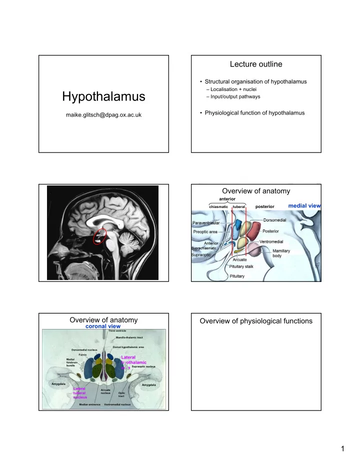
How does it all come together?
- Increased blood osmolarity causes osmosensitive (OVLT)
neurons to shrink
- TRPV1 channels open, leading to depolarisation and eventually
firing of (OVLT) neurons (graded response)
- (OVLT) neurons make monosynaptic glutamatergic contacts
with supra-optic nuclei neurons
- This promotes firing of ADH-releasing neurons and hence ADH
release
- ADH releasing neurons are intrinsically osmosensitive
Firing rate of ADH-releasing neurons depends on central and peripheral inputs as well as their intrinsic
ADH-release
How does it all come together?
- Increased blood osmolarity causes osmosensitive (OVLT)
neurons to shrink
- TRPV1 channels open, leading to depolarisation and eventually
firing of (OVLT) neurons (graded response)
- (OVLT) neurons make monosynaptic glutamatergic contacts
with supra-optic nuclei neurons
- This promotes firing of ADH-releasing neurons and hence ADH
release
- ADH releasing neurons are intrinsically osmosensitive
Firing rate of ADH-releasing neurons depends on central and peripheral inputs (baroreceptors!) as well as their intrinsic osmosensitivity – Central DI
ADH, resulting in excess urine output and dehydration
- following pituitary stalk
damage (accident)
produces no ADH
Diabetes insipidus Summary of neuroendocrine hypothalamus
GnRH (FSH, LH); GHRH (GH); DA (prolactin)
Reproduction; growth
CRH (ACTH); TRH (TSH); ADH;
Steroid hormone production, energy metabolism, water retention; social behaviours, reproduction
Sost (GH)
Growth
ADH; oxytocin
water retention; social behaviours, reproduction
Summary of neuroendocrine hypothalamus
GnRH (FSH, LH); GHRH (GH); DA (prolactin)
reproduction; energy metabolism
CRH (ACTH); TRH (TSH); ADH;
behaviours, energy metabolism, water retention (blood flow), reproduction
Sost (GH)
energy metabolism
ADH; oxytocin
water retention (blood flow), behaviours, reproduction
Non-endocrine control via the hypothalamus