
SLIDE 1
1
Henry Moon Lecture
Ovarian tumors in the young Robert H. Young MD I have nothing to disclose
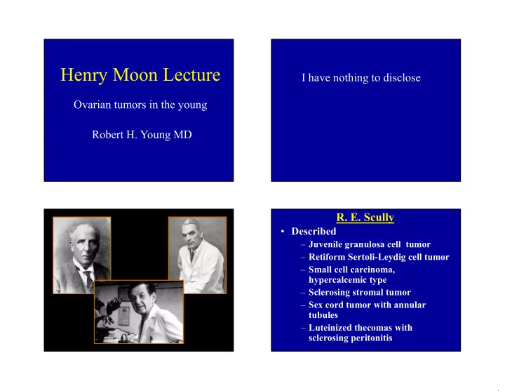
- R. E. Scully
- Described
– Juvenile granulosa cell tumor – Retiform Sertoli-Leydig cell tumor – Small cell carcinoma, hypercalcemic type – Sclerosing stromal tumor – Sex cord tumor with annular tubules – Luteinized thecomas with sclerosing peritonitis