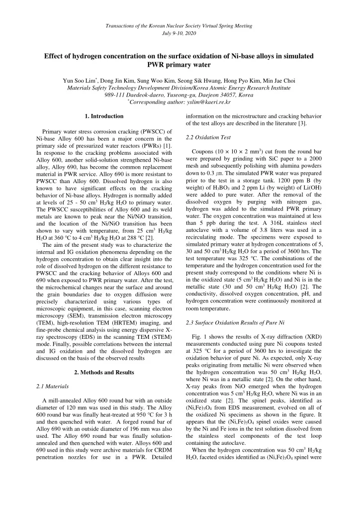

Transactions of the Korean Nuclear Society Virtual Spring Meeting July 9-10, 2020 Effect of hydrogen concentration on the surface oxidation of Ni-base alloys in simulated PWR primary water Yun Soo Lim * , Dong Jin Kim, Sung Woo Kim, Seong Sik Hwang, Hong Pyo Kim, Min Jae Choi Materials Safety Technology Development Division/Korea Atomic Energy Research Institute 989-111 Daedeok-daero, Yuseong-gu, Daejeon 34057, Korea * Corresponding author: yslim@kaeri.re.kr 1. Introduction information on the microstructure and cracking behavior of the test alloys are described in the literature [3]. Primary water stress corrosion cracking (PWSCC) of 2.2 Oxidation Test Ni-base Alloy 600 has been a major concern in the primary side of pressurized water reactors (PWRs) [1]. Coupons (10 × 10 × 2 mm 3 ) cut from the round bar In response to the cracking problems associated with were prepared by grinding with SiC paper to a 2000 Alloy 600, another solid-solution strengthened Ni-base mesh and subsequently polishing with alumina powders alloy, Alloy 690, has become the common replacement down to 0.3 ㎛ . The simulated PWR water was prepared material in PWR service. Alloy 690 is more resistant to prior to the test in a storage tank. 1200 ppm B (by PWSCC than Alloy 600. Dissolved hydrogen is also weight) of H 3 BO 3 and 2 ppm Li (by weight) of Li(OH) known to have significant effects on the cracking were added to pure water. After the removal of the behavior of Ni-base alloys. Hydrogen is normally added at levels of 25 - 50 cm 3 H 2 /kg H 2 O to primary water. dissolved oxygen by purging with nitrogen gas, hydrogen was added to the simulated PWR primary The PWSCC susceptibilities of Alloy 600 and its weld water. The oxygen concentration was maintained at less metals are known to peak near the Ni/NiO transition, than 5 ppb during the test. A 316L stainless steel and the location of the Ni/NiO transition has been shown to vary with temperature, from 25 cm 3 H 2 /kg autoclave with a volume of 3.8 liters was used in a H 2 O at 360 ℃ to 4 cm 3 H 2 /kg H 2 O at 288 ℃ [ 2]. recirculating mode. The specimens were exposed to simulated primary water at hydrogen concentrations of 5, The aim of the present study was to characterize the 30 and 50 cm 3 H 2 /kg H 2 O for a period of 3600 hrs. The internal and IG oxidation phenomena depending on the test temperature was 325 ℃ . The combinations of the hydrogen concentration to obtain clear insight into the temperature and the hydrogen concentration used for the role of dissolved hydrogen on the different resistance to present study correspond to the conditions where Ni is PWSCC and the cracking behavior of Alloys 600 and in the oxidized state (5 cm 3 H 2 /kg H 2 O) and Ni is in the 690 when exposed to PWR primary water. After the test, metallic state (30 and 50 cm 3 H 2 /kg H 2 O) [2]. The the microchemical changes near the surface and around conductivity, dissolved oxygen concentration, pH, and the grain boundaries due to oxygen diffusion were hydrogen concentration were continuously monitored at precisely characterized using various types of room temperature . microscopic equipment, in this case, scanning electron microscopy (SEM), transmission electron microscopy (TEM), high-resolution TEM (HRTEM) imaging, and 2.3 Surface Oxidation Results of Pure Ni fine-probe chemical analysis using energy dispersive X- Fig. 1 shows the results of X-ray diffraction (XRD) ray spectroscopy (EDS) in the scanning TEM (STEM) mode. Finally, possible correlations between the internal measurements conducted using pure Ni coupons tested at 325 ℃ for a period of 3600 hrs to investigate the and IG oxidation and the dissolved hydrogen are discussed on the basis of the observed results oxidation behavior of pure Ni. As expected, only X-ray peaks originating from metallic Ni were observed when the hydrogen concentration was 50 cm 3 H 2 /kg H 2 O, 2. Methods and Results where Ni was in a metallic state [2]. On the other hand, 2.1 Materials X-ray peaks from NiO emerged when the hydrogen concentration was 5 cm 3 H 2 /kg H 2 O, where Ni was in an A mill-annealed Alloy 600 round bar with an outside oxidized state [2]. The spinel peaks, identified as diameter of 120 mm was used in this study. The Alloy (Ni,Fe) 3 O 4 from EDS measurement, evolved on all of 600 round bar was finally heat-treated a t 950 ℃ for 3 h the oxidized Ni specimens as shown in the figure. It and then quenched with water. A forged round bar of appears that the (Ni,Fe) 3 O 4 spinel oxides were caused Alloy 690 with an outside diameter of 196 mm was also by the Ni and Fe ions in the test solution dissolved from used. The Alloy 690 round bar was finally solution- the stainless steel components of the test loop annealed and then quenched with water. Alloys 600 and containing the autoclave. When the hydrogen concentration was 50 cm 3 H 2 /kg 690 used in this study were archive materials for CRDM penetration nozzles for use in a PWR. Detailed H 2 O, faceted oxides identified as (Ni,Fe) 3 O 4 spinel were
Transactions of the Korean Nuclear Society Virtual Spring Meeting July 9-10, 2020 found to randomly distribute on the surface with a a metallic state at this hydrogen concentration. The variety of sizes. On the other hand, when the hydrogen noticeable difference in IG oxidation between Fig. (a) concentration was 5 cm 3 H 2 /kg H 2 O, two types of oxides and (b) is that, while Ni was enriched in the oxidized emerged. First, most of the oxides on the surface had grain boundary when Ni was in a reduced state at a hydrogen concentration of 30 cm 3 H 2 /kg H 2 O, Ni pyramidal shapes and they were identified as NiO from Fig. 1. NiO oxides form in this case since the Ni reacted with diffusing oxygen, resulting in the formation specimen is in an oxidation state at this hydrogen of Ni-rich oxides in the oxidized grain boundary when concentration. Another type of oxide was found over the Ni was in an oxidized state at a hydrogen concentration of 5 cm 3 H 2 /kg H 2 O. NiO pyramidal oxides, and was identified as (Ni,Fe) 3 O 4 . Fig. 1 Results of XRD measurements of pure Ni at hydrogen concentrations of 50 and 5 cm 3 H 2 /kg H 2 O tested at 325 ℃ for (a) a period of 3600 hrs. 2.4 Surface Oxidation Results of Alloy 600 The electron microscopic results of Alloys 600 and 690 obtained at the hydrogen concentration of 50 cm 3 H 2 /kg H 2 O were similar to those at the hydrogen concentration of 30 cm 3 H 2 /kg H 2 O, respectively. Therefore, results obtained at the hydrogen concentrations of 5 and 30 cm 3 H 2 /kg H 2 O will be presented in this study. Fig. 2 was obtained from Alloy 600 at a hydrogen concentration of 30 and 5 cm 3 H 2 /kg H 2 O tested at 325 ℃ for a period of 3600 hrs, respectively, presenting (b) Fig. 2 STEM image and EDS spectrum images of O, Cr, Fe, a STEM image and EDS spectrum images of O, Cr, Fe, and Ni in Alloy 600 at hydrogen concentrations of (a) 30 cm 3 and Ni obtained using the K α 1 lines. A grain boundary and (b) 5 cm 3 H 2 /kg H 2 O, tested at 325 ℃ for 3600 hrs. is visible in the STEM image. In the O spectrum image, a continuous thin Cr-rich oxide layer appears to emerge. 2.5 Surface Oxidation Results of Alloy 690 The faceted oxides over the surface grain boundary were identified as (Ni,Fe) spinel. The most intriguing Fig. 3 shows a STEM image and EDS spectrum feature in Fig. 2 is that oxygen diffused down along the images of O, Cr, Fe and Ni around the surface obtained grain boundary, causing the grain boundary to be from Alloy 690 at a hydrogen concentration of 30 and 5 oxidized irrespective of the hydrogen concentration. cm 3 H 2 /kg H 2 O, respectively, tested at 325 ℃ for a On the oxidized grain boundary, Cr was enriched. period of 3600 hrs. The surface oxidation layers were However, Fe and Ni were depleted in the oxidized grain much thicker than those of Alloy 600 under the same boundary (Fe and Ni spectrum images) compared to the experimental conditions. The most noticeable feature in average concentration of the matrix. This indicates that this figure is that there is no oxygen diffusion along a Cr oxide formed in the oxidized grain boundary. Ni was grain boundary which indicates that IG oxidation did enriched inside the oxidized grain boundary near the not occur in Alloy 690 irrespective of the hydrogen surface in the case of Fig. 2(a), which was obtained at concentration, in contrast with Alloy 600. From these the hydrogen concentration of 30 cm 3 H 2 /kg H 2 O. The results, it can be confirmed that the internal and IG formation of Ni-enriched regions is very common oxidation phenomena of Alloy 690 were quite different during the IG oxidation process of Alloy 600 in primary from those of Alloy 600. The main reason for these water whenever oxygen penetrates the metal and Cr is differences is thought to be the different chemical internally oxidized to form an oxide [4]. This behavior compositions, especially the different Cr contents in of Ni is thought to be mainly attributable to Ni being in
Recommend
More recommend