
SLIDE 1
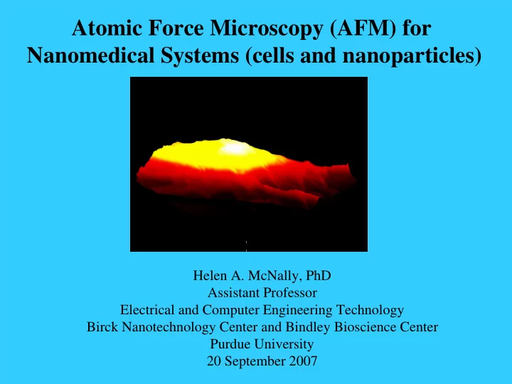
Atomic Force Microscopy (AFM) for Nanomedical Systems (cells and nanoparticles)
Helen A. McNally, PhD Assistant Professor Electrical and Computer Engineering Technology Birck Nanotechnology Center and Bindley Bioscience Center Purdue University 20 September 2007