SLIDE 6 8/15/2015 6
AFM image of peptide amphiphiles fibers AFM image of dip-pen pattern of peptide amphiphiles fibers, scale: 10um, by courtesy of H. Jiang
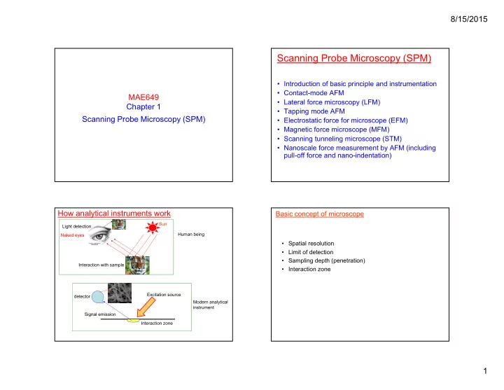
Application of AFM and SIMS in biology
SIMS image of peptide amphiphiles pattern CN-
Lateral force microscopy (LFM) images
- r LFM, the probe is scanned sidewise, and the friction
signal is calculated. The degree of torsion of the cantilever supporting the probe is a relative measure of surface friction caused by the lateral force exerted on the scanning probe. Note that for contact mode, the deflection signal is calculated as laser spot intensity for quadrants (A + B) - (C +D). Topographic (left) and LFM (right) images of a natural rubber/EDPM
- blend. 12μm scans, by M.G.
Heaton, et al Topographic (left) and LFM (right) images of the surface of a polished polycrystalline silicon carbide film. The polishing process
- bscures in the topography image the grain
structure, which is clearly visible in the LFM
- image. 30μm scans. by M.G. Heaton, et al
Distinguish the different phases in materials by LFM
Lateral force microscopy (LFM) images
A specialized use of LFM is Chemical Force Microscopy (CFM), where the tip is functionalized with a chemical species, and scanned over a sample to detect adhesion differences between the species on the tip and those on the surface
(1). CFM scan of well-defined regions that terminate in either methyl or carboxylic acid groups. (2). When a carboxylic acid-terminated tip is used for imaging (left), the carboxylic acid terminated regions exhibit greater frictional force (lighter color) than the methyl- terminated regions. (3). When a methyl terminated tip is used (right), the friction contrast is reversed. No differences are revealed by the topographic AFM scan (not shown) since the functional groups are structurally quite similar. (50μm images, Image courtesy of Dr.
- C. Lieber, Harvard University).
Broadening of features by tip
- The shape traced by the tip is in essence a superposition of spheres (neglecting
mechanical deformation, a later topic).
- Imaged lateral size is much larger than true size.
- Vertical size is approximately correct.
- Independent measurement of sphere size (e.g., via electron microscopy) or
distribution of sphere sizes (e.g., via scattering) can provide a calibration specimen: a means of determining the true shape of the tip via a nanoparticle.
Simple formulas describing the apparent width of objects