
SLIDE 1
Surgical Treatment of AVMs
Michael T. Lawton, MD
Chief, Vascular Neurosurgery Professor and Vice-Chairman Tong-Po Kan Endowed Chair University of California - San Francisco
Michael T. Lawton, MD
Chief, Vascular Neurosurgery Professor and Vice-Chairman Tong-Po Kan Endowed Chair University of California - San Francisco
Disclosures
- Mizuho America, Inc.: Royalties
- Stryker: Consultant
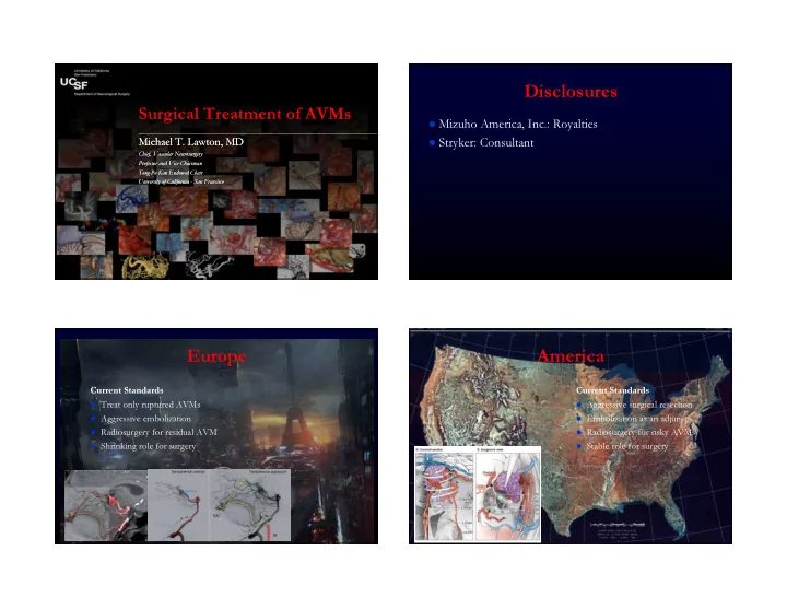
Europe
Current Standards
- Treat only ruptured AVMs
- Aggressive embolization
- Radiosurgery for residual AVM
- Shrinking role for surgery
America
Current Standards
- Aggressive surgical resection
- Embolization as an adjunct
- Radiosurgery for risky AVMs
- Stable role for surgery