
SLIDE 1
1
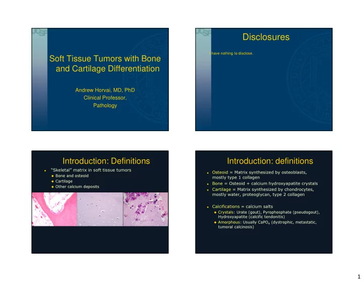
Soft Tissue Tumors with Bone and Cartilage Differentiation
Andrew Horvai, MD, PhD Clinical Professor, Pathology
Disclosures
I have nothing to disclose.
Introduction: Definitions
- “Skeletal” matrix in soft tissue tumors
Bone and osteoid Cartilage Other calcium deposits
Introduction: definitions
- Osteoid = Matrix synthesized by osteoblasts,
mostly type 1 collagen
- Bone = Osteoid + calcium hydroxyapatite crystals
- Cartilage = Matrix synthesized by chondrocytes,
mostly water, proteoglycan, type 2 collagen
- Calcifications = calcium salts
Crystals: Urate (gout), Pyrophosphate (pseudogout),
Hydroxyapatite (calcific tendonitis)
Amorphous: Usually CaPO4 (dystrophic, metastatic,
tumoral calcinosis)