
SLIDE 1
5/25/19 1
Bone Pathology for the Surgical Pathologist
UCSF Current Issues in Pathology 2019
Andrew Horvai MD PhD Clinical Professor, Pathology UCSF, San Francisco, CA
Disclosure
Company Relationship type Presage Biosciences Consultant
Outline
- Approach to bone pathology
- Decalcification
- Osteomyelitis
- Avascular necrosis
- Infected arthroplasty
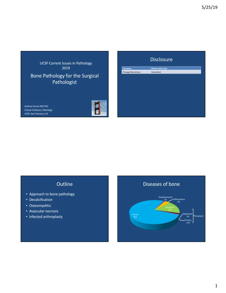
Diseases of bone
Trauma 76% Developmental 1% Inflammatory 4% Metabolic 17% Metastatic 1% Primary <1% Neoplasm