SLIDE 12 3/26/2014 12
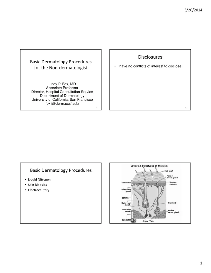
Where to Biopsy
Lesion Location of biopsy Tumor Thickest portion, avoid necrotic tissue Blister Edge of the lesion, include about 2mm
- f blister edge; send for H+E and DIF
Ulceration/necrotic lesion Edge of ulcer or necrosis plus adjacent skin Generalized polymorphic eruption Characteristic lesion of recent onset (+/‐ more developed lesion) Small vessel vasculitis (palpable purpura) Characteristic lesion of recent onset (ideally <24 hours old)
Adapted from: Bolognia, Jorizzo, and Schaffer. Dermatology 3rd ed. Elsevier 2012
Direct Immunofluorescence
- Location of the biopsy depends on diagnosis
- Vasculitis‐ lesional skin from an early lesion
- Lupus
– DLE/SCLE Lesional skin – SLE‐ Lesional, uninvolved can be positive as wel
– Peri‐lesional
Slide courtesy of Jeff North, MD Slide courtesy of Jeff North, MD
DIF‐ peri‐lesional
- Eclipsing the edge of new
blister
- Being too far from a blister
can cause false negative DIF
DIF in Pemphigoid and Pemphigus
Slide courtesy of Jeff North, MD Photo courtesy of Kari Connolly, MD