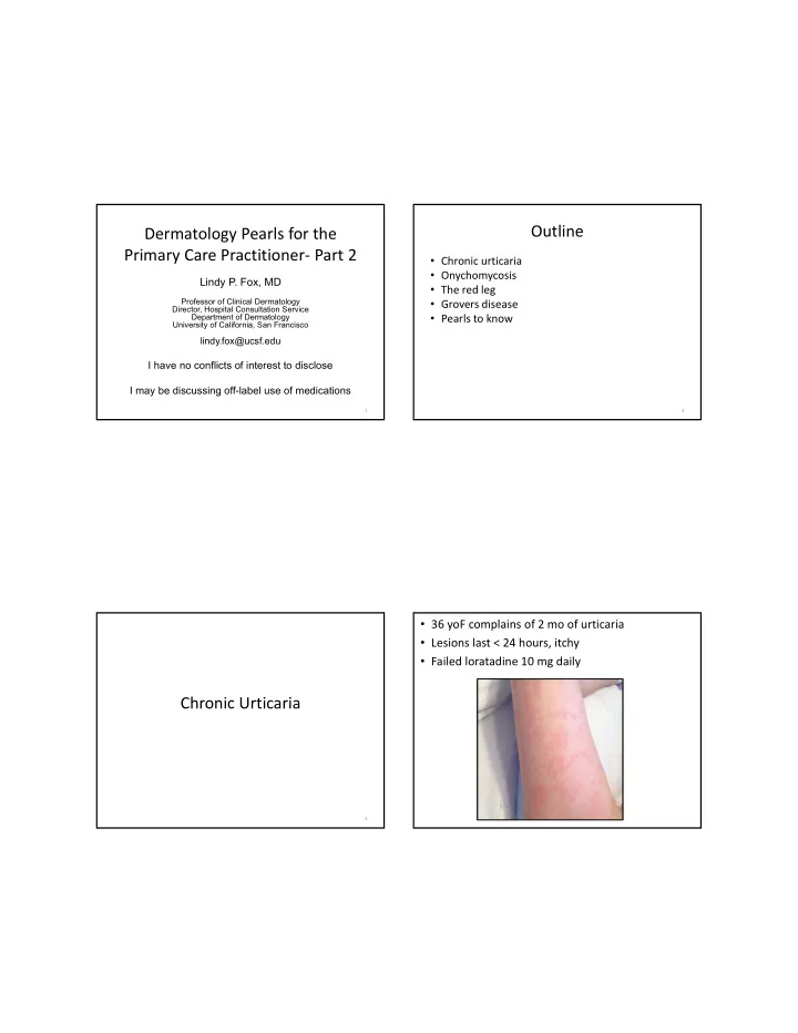
outline dermatology pearls for the primary care
play

Outline Dermatology Pearls for the Primary Care Practitioner Part 2 - PDF document

Outline Dermatology Pearls for the Primary Care Practitioner Part 2 Chronic urticaria Onychomycosis Lindy P. Fox, MD The red leg Professor of Clinical Dermatology Grovers disease Director, Hospital Consultation Service


  1. Outline Dermatology Pearls for the Primary Care Practitioner‐ Part 2 • Chronic urticaria • Onychomycosis Lindy P. Fox, MD • The red leg Professor of Clinical Dermatology • Grovers disease Director, Hospital Consultation Service • Pearls to know Department of Dermatology University of California, San Francisco lindy.fox@ucsf.edu I have no conflicts of interest to disclose I may be discussing off-label use of medications 1 2 • 36 yoF complains of 2 mo of urticaria • Lesions last < 24 hours, itchy • Failed loratadine 10 mg daily Chronic Urticaria 3

  2. Chronic Urticaria Chronic Urticaria‐ Workup • History and physical guides workup • Urticaria, with or without angioedema > 6 • Labs to check weeks – CBC with differential – Lesions last < 24 hours, itch, completely resolve – ESR, CRP • Divided into chronic spontaneous (66‐93%) – TSH and thyroid autoantibodies or chronic inducible – Liver function tests • Natural history‐ 2‐5 years – CU Index (Fc‐εRIα Ab or Ab to IgE) – Maybe tryptase for severe, chronic recalcitrant disease – > 5 yrs in 20% patients – 13% relapse rate – Maybe look for bullous pemphigoid in an older patient • Etiology • Provocation for inducible urticaria – 30 ‐50 % ‐ IgG autoAb to IgE or FcεRIα – Remainder, unclear Eur J Dermatol 2016 Clin Transl Allergy 2017. 7(1): 1‐10 Allergy Asthma Immunol Res. 2016;8(5):396‐403 Eur J Dermatol 2016 Clin Transl Allergy 2017. 7(1): 1‐10 J Allergey Clin Immunol Pract. 2017. Sept 6. S2213‐2198 Chronic Spontaneous Urticaria‐ Treatment First line H1 antihistamines‐ 2 nd generation What does my “second line” look like? <40% respond to standard dose H1 blockade Avoid triggers (NSAIDS, ASA) Can increase to up to 4X standard dose 60% chance of response Second line • Fexofenadine 360 mg am, 180 mg noon, 360 mg pm High dose 2 nd generation AH • Cetirizine 10 mg BID Add another 2 nd generation AH • Hydroxyzine 25 mg QHS 1 st gen H1 antihistamine QHS +/‐ H2 antagonist • +/‐ Monteleukast 10 mg QD +/‐ Leukotriene antagonist • +/‐ Ranitidine 300 mg QD Third line Omalizumab • Give epipens (3) Cyclosporine Dapsone Sulfasalazine Hydroxychloroquine J Allergy Clin Immunol 2014. 133(3):914‐5 BJD 2016. 175:1134–52 • When time to taper, take off 1 pill per week Mycophenolate mofetil Clin Transl Allergy 2017. 7(1): 1‐10 TNFα antagonists Allergy Asthma Immunol Res. 2016;8(5):396‐403 Eur J Dermatol 2016 (epub ahead of print) Anti CD20 Ab (rituximab) Allergy Asthma Immunol Res. 2017 November;9(6):477‐482. Allergy 2018. Jan 15. epub ahead of print

  3. CSU‐ when to refer • Atypical lesion morphology or symptoms – > 24 hours, central duskiness/purpura – Asymptomatic or burn >> itch Onychomycosis • Minimal response to medications – High dose H1 nonsedating antihistamines – H1 sedating antihistamines • Associated symptoms – Fever, fatigue, mylagias, arthralgias • Elevated ESR/CRP 10 Onychomycosis Onychomycosis • Infection of the nail plate by fungus • Vast majority are due to dermatophytes, especially Trichophyton rubrum • Very common • Increases with age • Half of nail dystrophies are onychomycosis • This means 50% of nail dystrophies are NOT fungal 11 12

  4. Onychomycosis Onychomycosis Diagnosis • KOH is the best test, as it is cheap, accurate if positive, and rapid; Positive 59% • If KOH is negative, perform a fungal culture • Frequent contaminant overgrowth • 53% positive • Nail clipping • Send to pathology lab to be sectioned and stained with special stains for fungus • Accurate (54% positive), rapid (<7d), written report • Downside: Cost (>$100) 13 14 Onychomycosis Onychomycosis: Local Treatment Interpreting Nail Cultures • Laser‐ insufficient data that it works • Any growth of T. rubrum is significant • Topical Therapy: • Contaminants • Ciclopirox (Penlac) 8% Lacquer: – Not considered relevant unless grown twice • Cure rates 30% to 35% for mild to moderate onychomycosis from independent samples AND no (20% to 65% involvement) dermatophyte is cultured • Clinical response about 65% – Relevant contaminants: • Efinaconazole (Jublia) 10%* • C. albicans • Daily for 48 weeks • Scopulariopsis brevicaulis • Complete or almost complete cure (completely clear nail)‐ 26% • • Fusarium Mycologic cure (neg KOH and neg fungal cx)‐ 55% • • Scytalidium (Carribean, Japan, Europe) Tavaborole (Kerydin) 5%* – Especially in immunosuppressed patients • Daily for 48 weeks • Complete or almost complete cure (completely clear nail)‐ 15‐17% • Mycologic cure (neg KOH and neg fungal cx)‐ 31‐36% 15 16 *Data from pharma website

  5. Onychomycosis Onychomycosis: Systemic Treatment Assessing Treatment Efficacy • • Nail growth Itraconazole: – At 2 to 3 months nail begins to grow out – 200 mg/d for 3 months – Continues for 12 months – 400 mg/d for one week per month for 4 months • Repeat KOH/culture at 4-6 months • Terbinafine: 250mg po QD – If culture still positive, treatment will likely fail – Fingernails: 6 weeks – KOH may still be positive (dead dermatophytes) – Toenails: 12 weeks • Failures • Check LFTs at 6 weeks – Pulse dosing – Terbinafine resistance • 500 mg daily for one week monthly for 3 months – Non-dermatophyte molds – Efficacy: 35% complete cures; 60% clinical cures – Dermatophytoma 17 18 The red leg: Cellulitis and its (common) mimics • Cellulitis/erysipelas • Stasis dermatitis • Contact dermatitis 19

  6. Cellulitis • Infection of the dermis • Gp A beta hemolytic strep and Staph aureus • Rapidly spreading • Erythematous, tender plaque, not fluctuant • Patient often toxic • WBC, LAD, streaking • Rarely bilateral • Treat tinea pedis

  7. Stasis Dermatitis Contact Dermatitis • Itch (no pain) • Often bilateral, L>R • Patient is non‐toxic • Itchy and/or painful • Red, hot, swollen leg • Erythema and • No fever, elevated WBC, edema can be severe LAD, streaking • Look for sharp cutoff • Look for: varicosities, • Treat with topical edema, venous ulceration, hemosiderin deposition steroids • Superimposed contact dermatitis common Contact Dermatitis • Common causes – Applied antibiotics (Neomycin, Bacitracin) – Topical anesthetics Grover’s Disease (benzocaine) – Other (Vitamin E, topical diphenhydramine) • Avoid topical antibiotics to leg ulcers – Metronidazole OK (prevents odor) 28

  8. Grovers Disease (transient acantholytic dermatosis) • Sudden eruption of papules, papulovesicles; often crusted • Mid chest and back • Itchy • Middle aged to older men • Etiology unknown‐ heat, sweating • Risk factors: hospitalized, febrile, sun damage • Transient • Treatment: topical steroids (triamcinolone 0.1% cream); get patient to move around Pearls to know

  9. Pustular Psoriasis • Pustular and erythrodermic variants of psoriasis • Can be life‐threatening • Most common in patients who carry a diagnosis of psoriasis who have been given systemic steroids and then tapered • High cardiac output state with risk of high output failure • Electrolyte imbalance (Ca 2+ ), respiratory distress, temperature dysregulation • Best treated with hospitalization and cyclosporine or acitretin 33 34 35 36

  10. Lotrisone Tinea Incognito • Combination of betamethasone plus clotrimazole – Weak antifungal + superpotent steroid • Inadequate to kill fungus and may cause complications (striae, fungal folliculitis) • Dermatologists rarely use it • Rarely indicated 37 38 Case • 67M underwent an elective saphenous vein phlebectomy for asymptomatic varicosities • 4d post op, he develops erythema around the wound. • Ulceration continues to expand despite multiple debridements and broad spectrum antibiotics. • Wound cultures are negative • 3 weeks later, he is transferred to UCSF and a dermatology consultation is called • Tmax 104, WBC 22

  11. Pyoderma Gangrenosum • Rapidly progressive (days) ulcerative process • Begins as a small pustule which breaks down forming an ulcer • Undermined violaceous border • Expands by small peripheral satellite ulcerations which merge with the central larger ulcer • Occur anywhere on body • Triggered by trauma (pathergy) (surgical debridement, attempts to graft) Pyoderma Gangrenosum Pyoderma Gangrenosum • 50% have no • Workup underlying cause – Skin biopsy for H&E and culture • Associations (50%): – Rheumatoid factor – Inflammatory bowel – SPEP/UPEP disease (1.5%-5% of – ANCA (ulcers of Granulomatosis with IBD patients get PG) Polynagiitis can mimic PG) – Rheumatoid arthritis – Seronegative arthritis – Colonscopy (r/o IBD) – Hematologic – Peripheral smear, Bone marrow biopsy (r/o abnormalities (AML) AML)

Download Presentation
Download Policy: The content available on the website is offered to you 'AS IS' for your personal information and use only. It cannot be commercialized, licensed, or distributed on other websites without prior consent from the author. To download a presentation, simply click this link. If you encounter any difficulties during the download process, it's possible that the publisher has removed the file from their server.

Recommend


More recommend