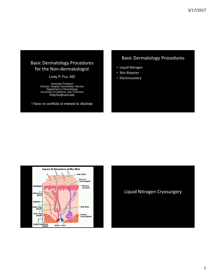
basic dermatology procedures basic dermatology procedures
play

Basic Dermatology Procedures Basic Dermatology Procedures Liquid - PDF document

3/17/2017 Basic Dermatology Procedures Basic Dermatology Procedures Liquid Nitrogen for the Non dermatologist Skin Biopsies Lindy P. Fox, MD Electrocautery Associate Professor Director, Hospital Consultation Service Department of


  1. 3/17/2017 Basic Dermatology Procedures Basic Dermatology Procedures • Liquid Nitrogen for the Non ‐ dermatologist • Skin Biopsies Lindy P. Fox, MD • Electrocautery Associate Professor Director, Hospital Consultation Service Department of Dermatology University of California, San Francisco lindy.fox@ucsf.edu I have no conflicts of interest to disclose 1 Liquid Nitrogen Cryosurgery 1

  2. 3/17/2017 Liquid Nitrogen Cryosurgery Liquid Nitrogen Cryosurgery Principles • ‐ 196°C ( − 320.8°F) • Indications • Temperatures of − 25°C to − 50°C ( − 13°F to − 58°F) – Benign, premalignant, in situ malignant lesions within 30 seconds with spray or probe • Objective – Selective tissue necrosis • Benign lesions: − 20°C to − 30°C ( − 4°F to − 22°F) • Reactions predictable • Malignant lesions: − 40°C to − 50°C. – Crust, bulla, exudate, edema, sloughing • Rapid cooling  intracellular ice crystals • Post procedure hypopigmentation • Slow thawing  tissue damage – Melanocytes are more sensitive to freezing than • Duration of THAW (not freeze) time is most keratinocytes important factor in determining success Am Fam Physician. 2004 May 15;69(10):2365 ‐ 2372 Liquid Nitrogen Cryosurgery • Fast freeze, slow thaw cycles – Times vary per condition (longer for deeper lesion) – One cycle for benign, premalignant – Two cycles for warts, malignant (not commonly done) • Lateral spread of freeze (indicates depth of freeze) – Benign lesions 1 ‐ 2mm beyond margins – Actinic keratoses ‐ 2 ‐ 3mm beyond margins – Malignant ‐ 3 ‐ 5+mm beyond margins (not commonly done) From: Bolognia, Jorizzo, and Schaffer. Dermatology 3 rd ed. Elsevier 2012 2

  3. 3/17/2017 Liquid Nitrogen Cryosurgery Cryosurgery for Common Warts Technique • Freeze time 20 ‐ 60 seconds • Hold spray gun 1 ‐ 1.5cm away from target • Margin ‐ 2 ‐ 3mm • Freeze until ice field fills the margin • Thaw 30 ‐ 45 seconds • Maintain the spray for the appropriate time • TWO cycles better than one BEYOND initial time of ice field formation • Repeat every 3 ‐ 4 weeks • If more than one cycle required, allow for • Average # of warts cleared= 40% complete thawing before beginning next cycle • Average # of treatments to clear warts = 12 – ONE YEAR! 3

  4. 3/17/2017 Cryosurgery for Planar Warts Cryosurgery for Actinic Keratoses • One freeze ‐ thaw cycle • margin ‐ 2 ‐ 3mm • May consider • Freeze time cotton tipped – AK 5 ‐ 7s applicator – Actinic cheilitis 10 ‐ 20s technique www.dermquest.com Cryosurgery for Lentigines Cryosurgery for Seborrheic Keratoses • Freeze ‐ thaw cycle • Quick 3 ‐ 4s freeze depends on thickness • Avoid overfreezing • Thin/flat ‐ freeze 5 ‐ 10s – Risk of hypopigmentation • Large/thick ‐ freeze >10s, may need second cycle 4

  5. 3/17/2017 Cryosurgery for SCC in situ * • One 30 second freeze Or • Two 20 second freezes Skin Biopsies • Close follow up *ED+C still preferred treatment option Skin Biopsy Skin Biopsy Types • Procedure itself is easy • Curettage • Knowing when and where to biopsy much • Snip/scissors more difficult • Shave biopsy • Pathologist can only comment on the tissue • Saucerization provided (not what’s left on patient) • Punch • Potential pitfalls in technique • Incisional • Excisional ( in toto ) 5

  6. 3/17/2017 Curettage with Biopsy • Samples epidermis only • Clinically benign lesions involving the epidermis – Verrucae (warts), seborrheic keratoses, actinic keratoses • Send pathology at same time as treating the lesion • Limitations • Hold like pencil – Limited to the epidermis • Draw pressure under the lesion (epidermis) – Fragmented tissue From: Bolognia, Jorizzo, and Schaffer. Dermatology 3 rd ed. Elsevier 2012 Snip/Scissors Biopsy • Pedunculated lesions • Benign growths – Acrochordons (skin tags) – Filiform warts – Pedunculated nevi • If very thin attachment to skin (stalk) don’t need anesthesia • Use iris or Gradle scissors • May require hemostasis with aluminum chloride, electrodesiccation From: Bolognia, Jorizzo, and Schaffer. Dermatology 3 rd ed. Elsevier 2012 6

  7. 3/17/2017 www.hovesskinclinic.co.uk Shave Biopsy • Samples epidermis and papillary (superficial) dermis • Ideal for elevated lesions involving the epidermis and superficial dermis – Inflammatory dermatoses of epidermis, superficial dermis (psoriasis, eczema, CTCL, lichen planus) – Nevi, benign adnexal tumors – Diagnosis of basal cell or squamous cell carcinoma – Diagnosis of lentigo maligna (MIS) Onsurg.com Am Fam Physician. 2011 Nov 1;84(9):995 ‐ 1002 Good Shave Biopsy • Be sure to get below simple hyperkeratosis and upper dermis • Palms, soles, hyperkeratotic lesions • Require hemostasis with aluminum chloride, electrodesiccation From: Bolognia, Jorizzo, and Schaffer. Dermatology 3 rd ed. Elsevier 2012 Slide courtesy of Jeff North, MD 7

  8. 3/17/2017 Saucerization Biopsy • Deeper biopsy with intentional deeper placement of the blade • Samples epidermis and superficial and deep dermis • Advantage – Histologic examination of the entire circumference of the lesion with adequate depth to assess invasion • Ideal for – Inflammatory dermatoses with dermal infiltrate • Intention is to get to deep dermis – Atypical pigmented lesions (to r/o melanoma) • Requires hemostasis with aluminum chloride, electrodesiccation – Keratoacanthoma/SCC From: Bolognia, Jorizzo, and Schaffer. Dermatology 3 rd ed. Elsevier 2012 Punch Biopsy Punch Biopsy • Samples epidermis, dermis and superficial subcutaneous fat • Varying barrel sizes ‐ 2mm ‐ 8mm • Ideal for – Inflammatory dermatoses with deep dermal infiltrate (lupus) – Infiltrative diseases (amyloid, sarcoid, lymphoma cutis) – Blistering diseases (pemphigus, pemphigoid) – Depressed lesions (scleroderma) • Limitations • Stabilize skin around punch with free hand – Only samples portion of larger lesion • Twist with firm downward pressure in one direction – Requires suture (>3mm) – Not ideal for subcutaneous lesions • Gently lift tissue with forceps at edge of epidermis (do not crush) • NO contraindications to punch biopsy other than avoiding • If plug not elevating, angle scissors downward to base bowel and brain • Try to make sure there is some fat at the base of the sample Slide courtesy of Wilson Liao, MD 8

  9. 3/17/2017 Good Punch Biopsy Incisional Biopsy • Samples epidermis, dermis, subcutaneous fat • Removes wedge from center or edge of lesion • Ideal for – Large tumors – Subtle diseases of connective tissue – Diseases of the fat (panniculitis) – Diseases of the fascia Slide courtesy of Jeff North, MD Excisional Biopsy • Samples epidermis, dermis, subcutaneous fat • Intended to be definitive treatment • Ideal for – Suspected invasive melanoma From: Bolognia, Jorizzo, and Schaffer. Dermatology 3 rd ed. Elsevier 2012 9

  10. 3/17/2017 Crush Artifact Skin Biopsies ‐ Potential Pitfalls • Crush artifact • Leaving part of tissue in punch tool • Multiple specimens, mislabeling Slide courtesy of Jeff North, MD Failure to Deliver Multiple Biopsy Specimens • Leaving part of the biopsy in the punch tool • Critically important to have an established protocol/routine to ensure the correct biopsy goes in the correct bottle Biopsy C A B Slide courtesy of Jeff North, MD Slide courtesy of Jeff North, MD 10

  11. 3/17/2017 Shave Biopsy Tray Video courtesy of Wilson Liao, MD Punch Biopsy Tray 11

  12. 3/17/2017 How to biopsy a specific lesion Where to Biopsy Lesion Type of biopsy Lesion Location of biopsy Papulosquamous (eczema, Shave or saucerization biopsy Tumor Thickest portion, avoid necrotic tissue psoriasis) Blister Edge of the lesion, include about 2mm r/o melanoma Saucerization or excisional biopsy of blister edge; send for H+E and DIF Ulceration/necrotic Edge of ulcer or necrosis plus adjacent Blister Punch biopsy at the edge for H+E and DIF lesion skin Generalized Characteristic lesion of recent onset (+/ ‐ Wart, seborrheic keratosis, Shave biopsy or curettage polymorphic eruption more developed lesion) actinic keratosis Small vessel vasculitis Characteristic lesion of recent onset Scalp (alopecia) Punch biopsy from hair containing region (palpable purpura) (ideally <24 hours old) adjacent to alopecia, request transverse sections Adapted from: Bolognia, Jorizzo, and Schaffer. Dermatology 3 rd ed. Elsevier 2012 Direct Immunofluorescence • Location of the biopsy depends on differential diagnosis • Michel’s medium (not formalin) • Vasculitis ‐ lesional skin from an early lesion • Lupus – DLE/SCLE Lesional skin – SLE ‐ Lesional, uninvolved can be positive as well • Blistering – Peri ‐ lesional Slide courtesy of Jeff North, MD Slide courtesy of Jeff North, MD 12

  13. 3/17/2017 DIF in Pemphigoid and Pemphigus DIF in Other Immunobullous Disease • Dermatitis herpetiformis DIF ‐ peri ‐ lesional • Up to 1 cm away from lesion • Eclipsing the edge of new • Don’t overlap the clinical lesion blister • Being too far from a blister • Higher risk for loss of epidermis and can cause false negative DIF destruction of Ig by the neutrophilic inflammatory infiltrate • Serology: anti ‐ transglutaminase and anti ‐ endomysium antibodies also helpful Slide courtesy of Jeff North, MD Slide courtesy of Jeff North, MD Photo courtesy of Kari Connolly, MD Electrosurgery • Electrodesiccation – Superficial tissue destruction Electrosurgery • Electrocoagulation – Deep tissue destruction • Electrosection – Cutting 13

Download Presentation
Download Policy: The content available on the website is offered to you 'AS IS' for your personal information and use only. It cannot be commercialized, licensed, or distributed on other websites without prior consent from the author. To download a presentation, simply click this link. If you encounter any difficulties during the download process, it's possible that the publisher has removed the file from their server.

Recommend


More recommend