
SLIDE 1
7/13/16 1
2016 Internal Medicine Board Review Dermatology
Nina Botto/ Kanade Shinkai, MD PhD/ Lindy Fox, MD Assistant Professor of Dermatology Department of Dermatology University of California, San Francisco
1
Conflict of Interest
I have no relevant conflicts to disclose.
2
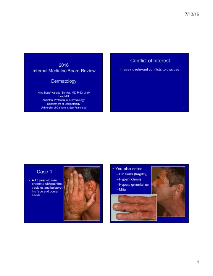
Case 1
- A 45 year old man
presents with painless vesicles and bullae on his face and dorsal hands
3
- You also notice
–Erosions (fragility) –Hypertrichosis –Hyperpigmentation –Milia
4