Conflict of Interest I have no relevant conflicts to disclose. 2 - - PDF document

conflict of interest
SMART_READER_LITE
LIVE PREVIEW

Conflict of Interest I have no relevant conflicts to disclose. 2 - - PDF document

7/1/2013 2013 Internal Medicine Board Review Dermatology Lindy Fox, MD Department of Dermatology University of California, San Francisco 1 Conflict of Interest I have no relevant conflicts to disclose. 2 Case 1 A 45 year old man


slide-1
SLIDE 1

7/1/2013 1 2013 Internal Medicine Board Review Dermatology

Lindy Fox, MD Department of Dermatology University of California, San Francisco

1

Conflict of Interest

I have no relevant conflicts to disclose.

2

Case 1

  • A 45 year old man

presents with painless vesicles and bullae on his face and dorsal hands

3

slide-2
SLIDE 2

7/1/2013 2

  • You also notice

– Erosions (fragility) – Hypertrichosis – Hyperpigmentation – Milia

4

Case 1, Question 1 The most likely diagnosis is:

  • A. Pemphigus vulgaris
  • B. Bullous impetigo
  • C. Bullous pemphigoid
  • D. Porphyria cutanea tarda
  • E. Dermatitis herpetiformis

5

Case 1, Question 1 The most likely diagnosis is:

  • A. Pemphigus vulgaris
  • B. Bullous impetigo
  • C. Bullous pemphigoid
  • D. Porphyria cutanea tarda
  • E. Dermatitis herpetiformis

6

slide-3
SLIDE 3

7/1/2013 3 Case 1, Question 2 Porphyria cutanea tarda

The underlying condition most likely to be associated is:

  • A. Hemochromatosis
  • B. Hepatitis C
  • C. Chronic renal insufficiency
  • D. Diabetes mellitus
  • E. NSAID use

7

Case 1, Question 2 Porphyria cutanea tarda

The underlying condition most likely to be associated is:

  • A. Hemochromatosis
  • B. Hepatitis C
  • C. Chronic renal insufficiency
  • D. Diabetes mellitus
  • E. NSAID use

8

Porphyria Cutanea Tarda (PCT)

  • Most common form of porphyria
  • Presents in 5th decade of life
  • M (60%), F (40%)
  • Risk factors

– HCV 85% – Hemochromatosis – Alcoholism – Genetic predisposition

  • Iron overload leads to reduced

uroporphyrinogen decarboxylase activity

9

slide-4
SLIDE 4

7/1/2013 4

Porphyria Cutanea Tarda

  • Sun-exposed sites (dorsal

hands, ears, face)

  • Non-inflammatory bulla
  • Skin fragility
  • Facial hypertrichosis
  • Milia
  • Hyperpigmentation

10

Porphyria Cutanea Tarda Treatment

  • Phlebotomy +/- erythropoetin
  • Low dose hydroxychloroquine

– 200 mg twice per week

  • Sun avoidance/photoprotection

11

Case 2

  • 43 yo Scandinavian male
  • Chronic abdominal pain,

diarrhea, weight loss

  • Small bowel biopsy:

shortening of intestinal villa

  • Pruritic papules and

vesicles on extensor surfaces and buttocks

  • No mucous membrane

involvement

12

slide-5
SLIDE 5

7/1/2013 5

13

Case 2, Question 1 The most likely diagnosis is:

  • A. Pemphigus vulgaris
  • B. Bullous impetigo
  • C. Bullous pemphigoid
  • D. Porphyria cutanea tarda
  • E. Dermatitis herpetiformis

14

Case 2, Question 1 The most likely diagnosis is:

  • A. Pemphigus vulgaris
  • B. Bullous impetigo
  • C. Bullous pemphigoid
  • D. Porphyria cutanea tarda
  • E. Dermatitis herpetiformis

15

slide-6
SLIDE 6

7/1/2013 6 Case 2, Question 2 Dermatitis Herpetiformis

This condition is most closely associated with:

  • A. Underlying lymphoma
  • B. Gluten-sensitive enteropathy
  • C. Autoimmune diseases
  • D. Diabetes mellitus
  • E. No associated underlying condition

16

Case 2, Question 2 Dermatitis Herpetiformis

This condition is most closely associated with:

  • A. Underlying lymphoma
  • B. Gluten-sensitive enteropathy
  • C. Autoimmune diseases
  • D. Diabetes mellitus
  • E. No associated underlying condition

17

Dermatitis Herpetiformis

  • Symmetric,

erythematous vesicles and papules in groups (“herpetiform”)

  • Intensely pruritic
  • Distribution is a clue:

– Elbows, knees, forearms, buttocks, scalp, neck

18

slide-7
SLIDE 7

7/1/2013 7 Dermatitis Herpetiformis Associated Diseases

  • Associated with gluten-sensitive enteropathy
  • Increased risk of GI lymphoma
  • Thyroid diseases in 20%

– hypothyroidism #1 – acute autoimmune thyroiditis – hyperthyroidism

  • Other: pernicious anemia, Addison’s disease

19

Dermatitis Herpetiformis

Diagnosis

20

Test Mode Result in DH Notes Skin biopsy H&E DIF Collections of neutrophils at dermal-epidermal junction DIF(+) granular IgA upper dermis IgA tissue transglutaminase ELISA, blood Sensitivity 90% Specificity 95% Higher false (+), confirm with anti- endomysial Ab IgA anti- endomysial Ab IF, blood (+) 70-90% Antigen is tissue transglutaminase

Dermatitis Herpetiformis

Treatment

  • Gluten free diet
  • Dapsone (50-300 mg daily) – rapid response
  • Does not respond to topical or systemic steroids

21

slide-8
SLIDE 8

7/1/2013 8

Pemphigus Vulgaris

  • Elderly
  • Widespread, friable

blisters -> erosions

  • Oral erosions
  • Paraneoplastic

association suggested by significant mucosal disease

22 23

Case 3

  • 48 yr old man
  • Facial rash x 3 months
  • Increasing fatigue
  • Difficulty stocking
  • verhead shelves

24

slide-9
SLIDE 9

7/1/2013 9

25 26 27

slide-10
SLIDE 10

7/1/2013 10
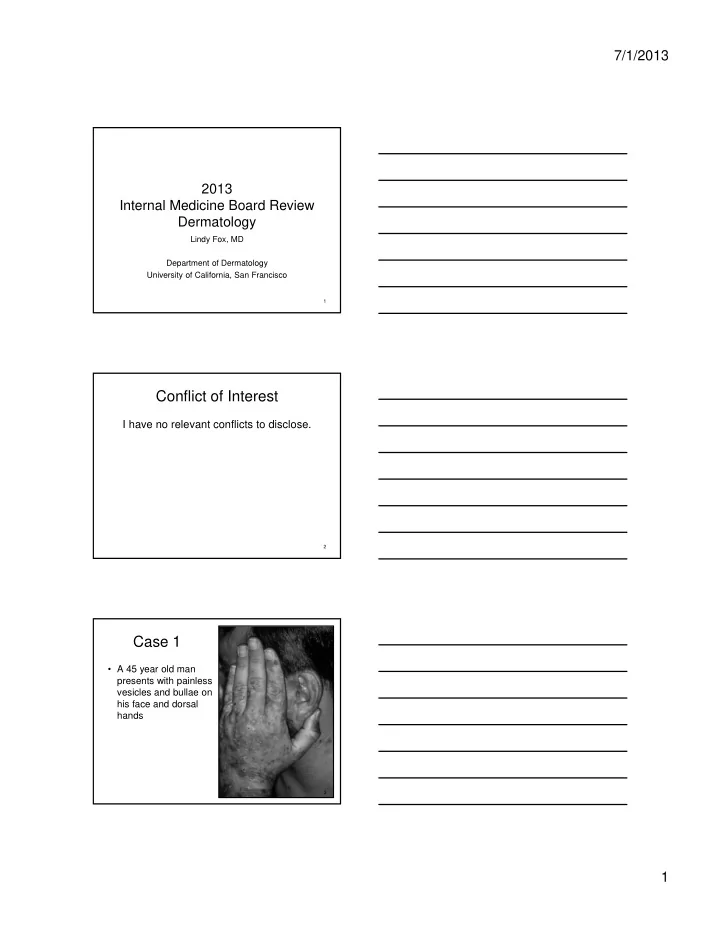
Case 3, Question 1

The lab test most likely to be abnormal is:

A. ESR B. Anti-smith antibody

  • C. Rheumatoid factor
  • D. Serum creatine kinase

E. Anti-dsDNA

28

Case 3, Question 1

The lab test most likely to be abnormal is:

A. ESR B. Anti-smith antibody

  • C. Rheumatoid factor
  • D. Serum creatine kinase

E. Anti-dsDNA

29

Dermatomyositis

  • Proximal muscle weakness
  • Characteristic skin findings

– Heliotrope: peri-orbital edema, violaceous rash @ eyelids – Gottron’s papules: flat, violaceous @ MCP, PIP, DIP joints – Photosensitive rash, shawl sign – Skin biopsy: similar to lupus (vacuolar interface + mucin)

  • Lab tests:

– Elevated CK or aldolase – Muscle biopsy, electromyography – ANA positive in 60-80% – Anti-Jo antibody associated with interstitial lung disease

30

slide-11
SLIDE 11

7/1/2013 11 Case 3, Question 2 Dermatomyositis

  • In an adult female patient with

dermatomyositis, which is the most important test to evaluate for an associated malignancy?

  • A. Thyroid scan
  • B. Mammogram
  • C. Colonoscopy
  • D. Upper endoscopy
  • E. Pelvic ultrasound

31

Case 3, Question 2 Dermatomyositis

  • In an adult female patient with

dermatomyositis, which is the most important test to evaluate for an associated malignancy?

  • A. Thyroid scan
  • B. Mammogram
  • C. Colonoscopy
  • D. Upper endoscopy
  • E. Pelvic ultrasound

32

Dermatomyositis Paraneoplastic Associations

  • Dermatomyositis is associated with

underlying malignancy in 32% of adult patients

– Risk highest > age 45, especially men

  • Women: ovarian cancer
  • Men: lung cancer
  • Asians: hepatomas, esophageal adenoCA

33

slide-12
SLIDE 12

7/1/2013 12

Case 4

  • Healthy 20 yo college student
  • Pruritic eruption x 10 days

34

Case 4, Question 1 The most likely diagnosis is:

  • A. Psoriasis
  • B. Pityriasis rosea
  • C. Secondary syphilis
  • D. Subacute cutaneous lupus
  • E. Tinea versicolor

35

Case 4, Question 1 The most likely diagnosis is:

  • A. Psoriasis
  • B. Pityriasis rosea
  • C. Secondary syphilis
  • D. Subacute cutaneous lupus
  • E. Tinea versicolor

36

slide-13
SLIDE 13

7/1/2013 13

Pityriasis Rosea

  • Common
  • Herald patch: 1 week

earlier, larger plaque

  • Widespread, symmetrical
  • val pink papules and

plaques with a central scale

  • Central trunk and back

(Christmas tree pattern)

  • Mimics the rash of

secondary syphilis

– CHECK RPR

37 38

Next case Subacute Cutaneous LE (SCLE)

  • Women aged 15-40
  • 50% meet ARA criteria for SLE, only 10%

severe

  • Renal or CNS disease rare = good prognosis
  • 80% ANA positive
  • Positive Ro/SSA

– Neonatal heart block is risk

  • Photosensitive

– Ro correlates with photosensitivity

39

slide-14
SLIDE 14

7/1/2013 14

Subacute Cutaneous LE Skin Lesions

  • Papulosquamous:

Resembles psoriasis

  • Annular
  • Sun-exposed areas
  • Face, V-neck chest,

and back

  • Heals without scarring
  • (unlike discoid LE)

40

Case 5

  • 55 yr old male
  • COPD, HTN, h/o psoriasis
  • Fever, shaking chills, and

diffuse erythema (erythroderma)

  • Meds:

– ACE inhibitor x 3 months – 1 week of pulsed prednisone with rapid taper for COPD flare

41 42

slide-15
SLIDE 15

7/1/2013 15 Case 5, Question 1 The most likely diagnosis is:

  • A. Drug eruption due to ACE inhibitor
  • B. Paraneoplastic syndrome due to non-small cell

lung cancer

  • C. Sézary syndrome (cutaneous T-cell

lymphoma)

  • D. Flare of psoriasis due to prednisone taper
  • E. Staphylococcal Scalded Skin Syndrome

43

Case 5, Question 1 The most likely diagnosis is:

  • A. Drug eruption due to ACE inhibitor
  • B. Paraneoplastic syndrome due to non-small cell

lung cancer

  • C. Sézary syndrome (cutaneous T-cell

lymphoma)

  • D. Flare of psoriasis due to prednisone taper
  • E. Staphylococcal Scalded Skin Syndrome

44

Pustular Psoriasis

  • Commonly drug-induced
  • Corticosteroid taper
  • Psoriasis flare + pustules
  • Can be life threatening

– High cardiac output state – Electrolyte imbalance – Respiratory distress – Temperature dysregulation

45

slide-16
SLIDE 16

7/1/2013 16

Psoriasis Comorbidities

  • Recent evidence links severe psoriasis with

– Arthritis – Cardiovascular disease (including MI) – Hypertension – Obesity – Diabetes – Metabolic syndrome – Malignancies

  • Lymphomas, SCCs, ? Solid organ malignancies

– Higher mortality – Poor quality of life

46 47

Psoriasis

48

slide-17
SLIDE 17

7/1/2013 17 Psoriasis Aggravators

  • Medications

– Systemic steroids – Beta blockers – Lithium – Hydroxychloroquine

  • Strep infections

– Guttate psoriasis (children)

  • Trauma
  • Sunburn
  • Severe life stress
  • HIV

– Up to 6% of patients with AIDS develop psoriasis

  • Alcohol for some
  • Smoking for some

49

Treatment for Psoriasis

  • Topical therapy

– Steroid ointment (start mid-potency) – Calcipotriene ointment – Tar

  • Phototherapy

– PUVA: psoralens + UVA

  • Systemic therapy

– Acitretin (oral retinoid) – Methotrexate, cyclosporine – Biologics

  • etanercept, infliximab, adalimumab (TNF alpha blockade)
  • ustekinumab (IL-12, IL-23 blockade)

**Systemic steroids are NOT on this list!**

50

Case 6

  • 42 yo HIV+ male admitted to ICU
  • Severely hypotensive  IV fluids, norepinephrine
  • ?Sepsis  antibiotics are started
  • History of taking TMP/SMX for UTI
  • 24 hrs after admission:
  • febrile
  • rash, rapidly progressive
  • skin is painful
  • gritty sensation in eyes
  • oral pain, difficulty swallowing

51

slide-18
SLIDE 18

7/1/2013 18

52

Case 6, Question 1 The most likely diagnosis is:

  • A. Drug Eruption
  • B. Staphylococcal Scalded Skin Syndrome
  • C. Autoimmune Blistering Disorder
  • D. Toxic Shock Syndrome
  • E. Severe viral exanthem

53

Case 6, Question 1 The most likely diagnosis is:

  • A. Drug Eruption
  • B. Staphylococcal Scalded Skin Syndrome
  • C. Autoimmune Blistering Disorder
  • D. Toxic Shock Syndrome
  • E. Severe viral exanthem

54

Toxic epidermal necrolysis (TEN)

slide-19
SLIDE 19

7/1/2013 19

Case 6, Question 2

All of the following are red flags of a serious drug eruption except:

  • A. Oral (mucocutaneous) involvement
  • B. Morbilliform eruption
  • C. Vesicle/ Bullae
  • D. Target lesions
  • E. Facial edema

55

Case 6, Question 2

All of the following are red flags of a serious drug eruption except:

  • A. Oral (mucocutaneous) involvement
  • B. Morbilliform eruption
  • C. Vesicle/ Bullae
  • D. Target lesions
  • E. Facial edema

56

Case 6, Question 3

All of the following are signs/symptoms of a drug hypersensitivity reaction except:

  • A. Eosinophilia
  • B. Lymphadenopathy
  • C. Elevated liver function tests
  • D. Hypertension
  • E. Facial edema

57

slide-20
SLIDE 20

7/1/2013 20

Case 6, Question 3

All of the following are signs/symptoms of a drug hypersensitivity reaction except:

  • A. Eosinophilia
  • B. Lymphadenopathy
  • C. Elevated liver function tests
  • D. Hypertension
  • E. Facial edema

58

Drug eruptions: Simple or Complex

Potentially life threatening Morbilliform drug eruption Drug hypersensitivity reaction Stevens-Johnson (SJS) Toxic epidermal necrolysis (TEN) Minimal systemic symptoms Systemic involvement

Simple Complex

59

Morbilliform (Simple) Drug Eruption

  • common
  • erythematous macules, papules
  • pruritus
  • no systemic symptoms
  • begins in 2nd week
  • risk factors: EBV, HIV infection
  • treatment:
  • D/C med if severe
  • symptomatic treatment:

diphenhydramine, topical steroids

60

slide-21
SLIDE 21

7/1/2013 21 Hypersensitivity Reactions Clinical features (General)

  • Rash
  • Fever
  • Pharyngitis
  • Hepatitis
  • Hematologic abnormalities

– eosinophilia – atypical lymphocytosis

  • Lymphadenopathy
  • Facial edema

61

Hypersensitivity Reactions

  • Skin eruption + visceral involvement
  • Late onset: 2- 6 weeks after medication started
  • Classic associations

– Aromatic anticonvulsants THESE CROSS-REACT

  • phenobarbital, carbamazepine, phenytoin, lamotrigine

ALSO – Allopurinol – Dapsone – NSAIDs – Sulfonamides – Anti-retrovirals

  • Treatment:

– Must stop medication (cannot treat through) – Prednisone (1-2mg/kg/day)

62

Anticonvulsant Hypersensitivity Reaction

63

Facial edema is key to the diagnosis of drug hypersensitivity reactions

slide-22
SLIDE 22

7/1/2013 22 Stevens-Johnson Syndrome (SJS)

Clinical morphology: – Target lesions – Two or more mucous membranes (eyes, mouth, genitalia) involved – Can progress to resemble toxic epidermal necrolysis (TEN)

64

Stevens-Johnson Syndrome (SJS)

65

Toxic Epidermal Necrolysis (TEN)

  • Life threatening
  • blistering reaction
  • Skin pain > pruritus
  • Erythema -> bullae ->

denuded dermis

  • Medical emergency: call

dermatology immediately

66

slide-23
SLIDE 23

7/1/2013 23

67

SJS/TEN: Emergency Management

  • Stop all unnecessary medications

– The major predictor of survival and severity of disease

  • Treatment

– Systemic

  • Check for Mycoplasma- 25% of SJS in pediatric

patients

  • Controversial

– SJS: high dose corticosteroids – TEN: IVIG 0.5-1g/kg/d x 4 days – Refer to burn unit or ICU early

  • Reduces risk of infection and reduces mortality to 5%
  • Call Ophthalmology

68

Case 7

  • 37 yo woman with inflammatory bowel disease
  • Rapidly progressive, painful ulceration
  • 3 days after bumping her leg on a chair

69

slide-24
SLIDE 24

7/1/2013 24

Case 7, Question 1

  • The most appropriate treatment for this

disorder is

  • A. Systemic steroids
  • B. Intravenous antibiotics
  • C. Surgical debridement
  • D. Compression dressing
  • E. Wet to dry dressings

70

Case 7, Question 1

  • The most appropriate treatment for this

disorder is

  • A. Systemic steroids
  • B. Intravenous antibiotics
  • C. Surgical debridement
  • D. Compression dressing
  • E. Wet to dry dressings

71

Pyoderma Gangrenosum

  • Rapidly progressive (days) ulcerative process
  • Begins as a small pustule
  • Undermined violaceous border
  • Occur anywhere on body
  • Triggered by trauma (pathergy) (surgical

debridement, attempts to graft)

72

slide-25
SLIDE 25

7/1/2013 25

Pyoderma Gangrenosum

  • 50% have no

underlying cause

  • Associations:

– Inflammatory bowel disease (1.5%-5% of IBD patients get PG) – Rheumatoid arthritis – Seronegative arthritis – Hematologic abnormalities

73

Pyoderma Gangrenosum Treatment

  • Treatment of underlying disease may not help PG
  • Topical therapy:

– Superpotent steroids – Topical tacrolimus

  • Systemic therapy:

– Systemic steroids – Cyclosporine – Tacrolimus – Mycophenolate mofetil – Thalidomide – TNF-inhibitors (infliximab)

74

Case 8

  • 52 yr old Caucasian

male

  • Wife noted enlarging

mole on his back

  • Family history –

brother recently diagnosed with melanoma, mother died of metastatic melanoma

75

slide-26
SLIDE 26

7/1/2013 26

Case 8,Question 1

The most significant prognostic indicator for melanoma is:

  • A. Anatomic location of melanoma
  • B. Diameter of melanoma
  • C. Positive family history in a first degree

relative

  • D. Thickness of melanoma
  • E. History numerous childhood blistering

sunburns

76

Case 8,Question 1

The most significant prognostic indicator for melanoma is:

  • A. Anatomic location of melanoma
  • B. Diameter of melanoma
  • C. Positive family history in a first degree

relative

  • D. Thickness of melanoma
  • E. History numerous childhood blistering

sunburns

77

Diagnosis of Melanoma

  • Prognosis is DEPENDENT on the depth of

lesion (Breslow’s depth) – < 1mm thickness is low risk – > 1mm Consider sentinel lymph node biopsy

  • If melanoma is on the differential, complete

excision or full thickness incisional biopsy is indicated

78

slide-27
SLIDE 27

7/1/2013 27

Malignant Melanoma

  • Asymmetry
  • Border
  • Color
  • Diameter (>6mm)
  • Evolution

79

Malignant Melanoma

80

Evolution

Case 9

  • 30 yr old Asian female
  • Fevers, malaise, arthralgia
  • Prior history of lower extremity

cellulitis with recent 10 day course of cephalexin

  • Meds: OCPs
  • ROS: intermittent abdominal

pain and loose stool

  • Family history: inflammatory

bowel disease

81

slide-28
SLIDE 28

7/1/2013 28

Case 9, Question 1

The most likely diagnosis is:

  • A. Erythema multiforme
  • B. Cellulitis resistant to cephalosporins
  • C. Pretibial myxedema
  • D. Erythema nodosum
  • E. Sweet’s syndrome

82

Case 9, Question 1

The most likely diagnosis is:

  • A. Erythema multiforme
  • B. Cellulitis resistant to cephalosporins
  • C. Pretibial myxedema
  • D. Erythema nodosum
  • E. Sweet’s syndrome

83

Case 9, Question 2

All of the following are associated with the erythema nodosum EXCEPT:

  • A. Oral contraceptive use
  • B. Streptococcal infection
  • C. Hyperthyroidism
  • D. Sarcoidosis
  • E. Inflammatory bowel disease

84

slide-29
SLIDE 29

7/1/2013 29

Case 9, Question 2

All of the following are associated with the erythema nodosum EXCEPT:

  • A. Oral contraceptive use
  • B. Streptococcal infection
  • C. Hyperthyroidism
  • D. Sarcoidosis
  • E. Inflammatory bowel disease

85

Erythema Nodosum: Associations

  • Common

– Idiopathic (35-55%) – Infection: STREP

  • URI, Mycoplasma, TB
  • Cocci (+ prognosis)

– Drugs

  • OCPs, sulfonamides,

PCN, halides – Sarcoidosis – IBD (Crohns > UC)

  • Uncommon

– Yersinia – Behçet's – Sweet’s syndrome – Pregnancy – Malignancy

  • Rare

– Brucellosis – Meninigococcus/gonococcus – E. coli – Pertussis – Syphilis – Leprosy – Cat Scratch – Chlamydia – Blastomycosis – Histoplasmosis – HIV

86

Erythema Nodosum Treatment

  • Elevation, bedrest, reduced exercise
  • Support stockings
  • NSAID’s or indomethacin (AVOID in

IBD)

  • SSKI (potassium iodide) 5-15 gtts TID

– Improvement can be seen within weeks

87

slide-30
SLIDE 30

7/1/2013 30

Case 10

  • 19 yr old male
  • Pruritic rash on left forearm
  • History of allergic rhinitis and

asthma

  • Meds: albuterol inhaler and

antihistamines prn

  • Rash improves with

triamcinolone ointment but recurs

88

Case 10, Question 1

Best next management step is:

  • A. Add antibiotic to cover bacterial

superinfection

  • B. Consider patch testing for contact allergen
  • C. Counsel patient on gentle skin care and use
  • f emollients
  • D. Add antiviral to cover for zoster
  • E. Treat for scabies

89

Case 10, Question 1

Best next management step is:

  • A. Add antibiotic to cover bacterial

superinfection

  • B. Consider patch testing for contact allergen
  • C. Counsel patient on gentle skin care and use
  • f emollients
  • D. Add antiviral to cover for zoster
  • E. Treat for scabies

90

slide-31
SLIDE 31

7/1/2013 31

Contact Dermatitis

  • Clinical Features

– Severe pruritus – Common allergens - poison oak, nickel, hair dye, bandages, topical neomycin, topical diphenhydramine – Worse in patients with atopic dermatitis

  • Diagnosis

– Look for clues as to shape and location – Linear (plant), wrist/beltline/earlobes (nickel), forehead/neck/ears (hair dye)

91

Treatment

  • For localized, small areas - potent topical

steroids and systemic anti-histamine

  • May need systemic steroids for 3 weeks to

prevent rebound reaction (ie poison oak)

92

Herpes Zoster

  • Hutchinson’s sign

– Vesicles on the nasal tip or side suggest nasociliary nerve branch involvement – prompt ophthalmology referral

  • Ramsay Hunt syndrome

– Vesicles in distribution of the nervus intermedius (external auditory canal, pinna, soft palate, anterior 2/3 of tongue) – Associated with vertigo, ipsilateral hearing loss, tinnitus, facial paresis

93

slide-32
SLIDE 32

7/1/2013 32

Treatment of Herpes Zoster

  • Early antiviral treatment (<3-4d)

– Faster resolution, shorter duration of pain – May help prevent and shorten duration of PHN

  • Prednisone

– Improves QOL score in patients with acute pain – Does not prevent PHN – Don’t use in immunosuppressed patients (increased risk of dissemination)

  • Post herpetic neuralgia (PHN)

– Capsaicin, amitriptyline, gabapentin

94

Case 11

  • 37 yo man with HCV

presents with fever, joint pain, and rash

  • Skin biopsy:

leukocytoclastic vasculitis

95

Case 11, Question 1

  • In this patient, the test most likely to be

abnormal is:

  • A. Antinuclear antibody
  • B. ASO
  • C. Cryoglobulins
  • D. Urinalysis
  • E. Stool guaiac

96

slide-33
SLIDE 33

7/1/2013 33

Case 11, Question 1

  • In this patient, the test most likely to be

abnormal is:

  • A. Antinuclear antibody
  • B. ASO
  • C. Cryoglobulins
  • D. Urinalysis
  • E. Stool guaiac

97

Leukocytoclastic Vasculitis Differential Diagnosis

  • Infection

– Post strep GN – Hepatitis B – SBE

  • Hypersensitivity

– Henoch-Schönlein purpura – Serum sickness – Medication

  • Rheumatic disease

– RA – Sjögren’s syndrome

  • Mixed cyroglobulinemia

(HCV)

  • Malignancy associated

– CLL – Multiple myeloma – Lymphoma – Hodgkin’s disease

  • ANCA associated

vasculitis

– Wegeners granulomatosis – Microscopic polyangiitis – Churg Strauss

99

slide-34
SLIDE 34

7/1/2013 34

Skin Cancer: Case 12

  • 65 year old man s/p

renal transplant

  • Rapid growth of nodule
  • n leg
  • Skin biopsy: Squamous

Cell Carcinoma

Squamous Cell Carcinoma

  • Red nodule (>1cm) or

tumor, often ulcerated or wart-like

  • Risk factors:

– Chronic sun exposure – Organ transplant recipients

101

Skin Cancer in Organ Transplant Recipients

  • Skin cancer is the most common malignancy
  • 90% are nonmelanoma skin cancer

– SCC: 70x increased risk – BCC: 10-20x increased risk – Melanoma: 2x increased risk

  • 2% of all deaths in OTR = metastatic SCC
  • Very aggressive tumors
  • Low threshold for skin biopsy in this patient

population

Traywick and O’Rielly. Derm Therapy. 2005; 18: 12-18

102

slide-35
SLIDE 35

7/1/2013 35 Skin Cancer in Organ Transplant Recipients

  • To reduce skin cancer risk in transplants:

– Reduce total immunosuppressive dose to minimum required – Absolute sun protection – Oral acitretin (25 mg daily) may reduce rate of SCC development

  • Refer organ transplant patients to a

dermatologist for regular skin checks

Good Luck!