Clinical characteristics Skin Cancer Precancerous lesions Common - - PDF document

clinical characteristics
SMART_READER_LITE
LIVE PREVIEW

Clinical characteristics Skin Cancer Precancerous lesions Common - - PDF document

Skin Caner Clinical characteristics Skin Cancer Precancerous lesions Common skin cancers Fernando Vega, MD Seattle Healing Arts ACTINIC KERATOSIS Common sun-induced Precancerous skin lesions premalignant neoplasm of the


slide-1
SLIDE 1

Skin Caner Fernando Vega, M.D. 1

Skin Cancer

Fernando Vega, MD Seattle Healing Arts Precancerous lesions Common skin cancers

Clinical characteristics

Precancerous skin lesions

Actinic keratoses Dysplastic melanocytic nevi
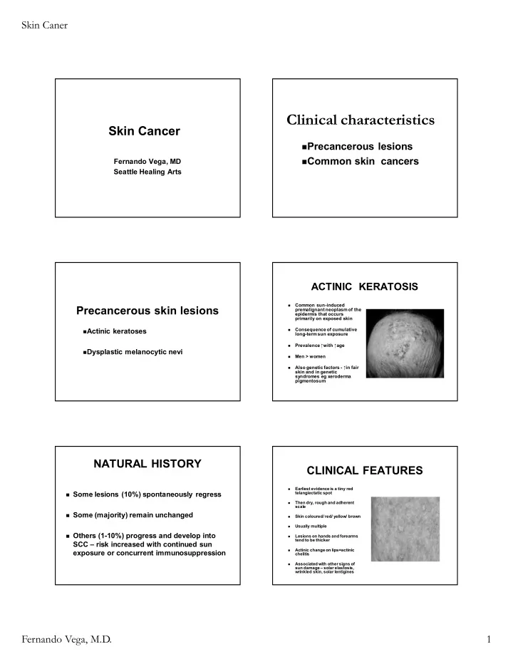
ACTINIC KERATOSIS

  • Common sun-induced

premalignant neoplasm of the epidermis that occurs primarily on exposed skin

  • Consequence of cumulative

long-term sun exposure

  • Prevalence ↑with ↑age
  • Men > women
  • Also genetic factors - ↑in fair

skin and in genetic syndromes eg xeroderma pigmentosum

NATURAL HISTORY

Some lesions (10%) spontaneously regress Some (majority) remain unchanged Others (1-10%) progress and develop into

SCC – risk increased with continued sun exposure or concurrent immunosuppression

CLINICAL FEATURES

  • Earliest evidence is a tiny red

telangiectatic spot

  • Then dry, rough and adherent

scale

  • Skin coloured/ red/ yellow/ brown
  • Usually multiple
  • Lesions on hands and forearms

tend to be thicker

  • Actinic change on lips=actinic

chelitis

  • Associated with other signs of

sun damage – solar elastosis, wrinkled skin, solar lentigines

slide-2
SLIDE 2

Skin Caner Fernando Vega, M.D. 2 Actinic keratoses Actinic keratoses

Actinic keratoses and SCC Actinic keratoses and SCC Actinic keratoses and BCC

Actinic keratoses

10% risk of malignant transformation

slide-3
SLIDE 3

Skin Caner Fernando Vega, M.D. 3 Hypertrophic AK’s Actinic cheilitis

Liquid nitrogen cryotherapy Topical therapies 5-FU (Efudex) Imiquimod (Aldara) Curettage for hypertrophic lesions

Treatment of AK’s

Residual hypopigmentation Blister formation

Liquid nitrogen Cryotherapy

Topical therapies

Efudex or Aldara

* 3-5 times per week * 6-8 weeks

Dysplastic nevi

  • Precursors for melanoma
  • When to biopsy
slide-4
SLIDE 4

Skin Caner Fernando Vega, M.D. 4

Miller A and M ihm M. N Engl J Med 2006;355:51-65

Biologic Events and Molecular Changes in the Progression of Melanoma

Tsao H et al. N Engl J Med 2004;351:998-1012

Clinical Images of Pigmented Lesions

Non-melanoma skin cancers

Basal cell carcinoma Squamous cell carcinoma Keratoacanthoma

Risk factors for development of BCC and SCC

Fair skin (Fitzpatrick’s types I-III) Blue eyes Red hair Family history Genetic syndromes Chronic sun exposure Old age Arsenic, tar

Basal cell carcinoma

BCC- clinical types

Nodular Superficial Morpheaform

slide-5
SLIDE 5

Skin Caner Fernando Vega, M.D. 5 Nodular BCC

Chronic lesion Easy bleeding Pearly border Surface telangiectasias Head and neck, trunk,

and extremities

slide-6
SLIDE 6

Skin Caner Fernando Vega, M.D. 6 Superficial BCC

Erythematous scaly

plaque

Slow growth Asymptomatic Trunk, extremities, face

Superficial BCC Morpheaform BCC

Resembles scar Asymptomatic and slow

growing

Ill-defined margins Marked subclinical

extension

BCC is the most

frequent skin cancer (80%)

BCC is 4x more

frequent than SCC

Metastases are rare

(<1% of cases)

Local destruction of

tissue

Treatment of BCC

Curettage electrodessication (ED/C) Surgical excision Traditional Mohs surgery Radiation therapy Topical therapy imiquimod

95% Cure Rate 50-75% Cure Rate

slide-7
SLIDE 7

Skin Caner Fernando Vega, M.D. 7

Squamous cell carcinoma

SCC types

In-situ Bowen’s disease Erythroplasia of Queyrat Invasive SCC Keratoacanthoma

Bowen’s disease

In-situ SCC Arsenic, HPV 16,

radiation

Invasive SCC

Erythematous nodule Indurated lesion Sun-exposed skin Men > women Slow growth

Invasive SCC Keratoacanthoma

Low grade SCC Rapid growth over

weeks

Trauma, sun exposure,

HPV 11 and 16

May progress to

invasive SCC

slide-8
SLIDE 8

Skin Caner Fernando Vega, M.D. 8

SCC is locally invasive and

destructive

Metastases in 1-3% of

cases

To lymph nodes 50-73% survival Distant sites (lungs) Incurable

Malignant Melanoma

Risk factors

Fair skin, red hair, and blue eyes Intermittent sun exposure Sunburns Tanning beds Freckles and melanocytic nevi Family history of melanoma

Clinical types- MM

Superficial spreading melanoma Lentigo maligna melanoma Acral lentiginous melanoma Nodular melanoma

ABCD of Melanoma

Asymmetry Border irregularity Color variegation Diameter >6mm

Tsao H et al. N Engl J Med 2004;351:998-1012

Clinical Images of Pigmented Lesions

slide-9
SLIDE 9

Skin Caner Fernando Vega, M.D. 9

Miller A and M ihm M. N Engl J Med 2006;355:51-65

Biologic Events and Molecular Changes in the Progression of Melanoma

Benign Melanocytic Neoplasms Benign Melanocytic Neoplasms Benign Melanocytic Neoplasms Benign Melanocytic Neoplasms

Congenital nevus

slide-10
SLIDE 10

Skin Caner Fernando Vega, M.D. 10 Malignant Melanoma Malignant Melanoma

With Regression

Malignant Melanoma

Supeerficial Spreading

Malignant Melanoma Malignant Melanoma

Ciliary Body

Malignant Melanoma

slide-11
SLIDE 11

Skin Caner Fernando Vega, M.D. 11 Malignant Melanoma Malignant Melanoma Malignant Melanoma Malignant Melanoma Malignant Melanoma Malignant Melanoma

slide-12
SLIDE 12

Skin Caner Fernando Vega, M.D. 12 Malignant Melanoma Malignant Melanoma Malignant Melanoma Malignant Melanoma Malignant Melanoma LENTIGO MALIGNA

An in situ pattern

  • f malignant

melanoma

Often reaches a

large size before the diagnosis is made

Lentigo → lentigo

maligna →lentigo maligna melanoma

slide-13
SLIDE 13

Skin Caner Fernando Vega, M.D. 13 CLINICAL FEATURES

Begins as a flat

pigmented lesion

Usually on sun-

exposed skin of head and neck

With time the colour

and border become more irregular

MANAGEMENT

Surgery – excision with a wide margin Radiotherapy Cryotherapy (deviation from rule) Immiquimod (by report)

Prognostic features- MM

Good prognosis Breslow < 1mm Intermediate prognosis Breslow 1-4mm Bad prognosis Breslow >4mm