
SLIDE 1
5/25/2017 1
Yunn-Yi Chen, MD, PhD Professor Director of Immunohistochemistry Laboratory UCSF
The Double-Edged Sword of Immunostains in Diagnostic Breast Pathology
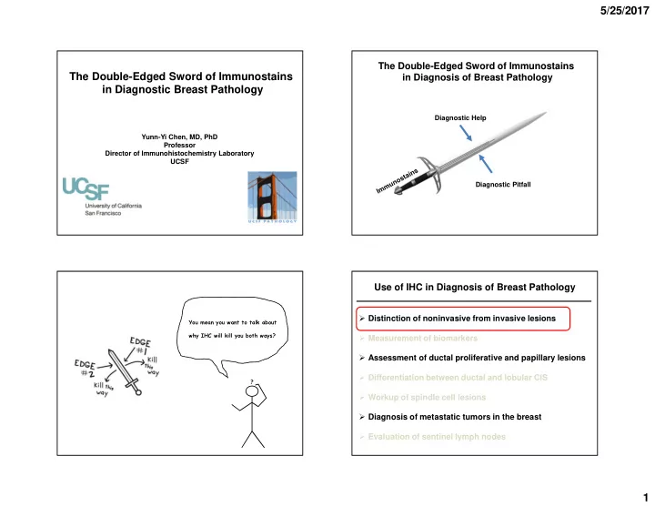
The Double-Edged Sword of Immunostains in Diagnosis of Breast Pathology
Diagnostic Help Diagnostic Pitfall
You mean you want to talk about why IHC will kill you both ways?