
4/2/2015 1
X-ray Diffraction (continued)
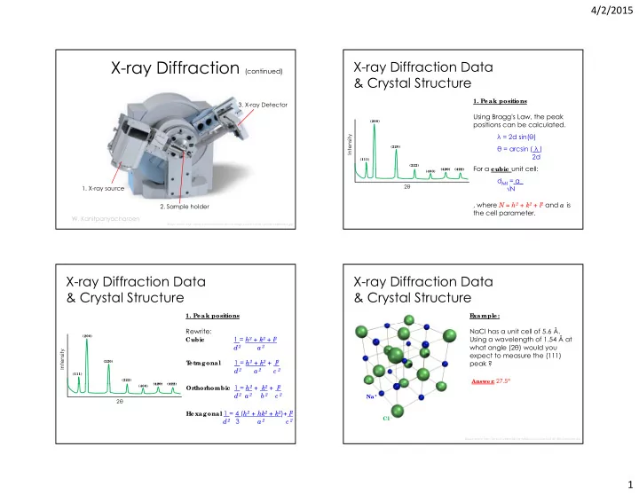
- 3. X-ray Detector
- W. Kanitpanyacharoen
I mage so urc e http:/ / www.h-and-m-analytic al.c o m/ image s/ se rvic e s/ x-ray-po wde r-diffrac tio n.jpg
- 1. X-ray source
- 2. Sample holder
X-ray Diffraction Data & Crystal Structure
- 1. Peak positions
(111) (200) (220) (222) (420) (422)
Intensity
Using Bragg's Law, the peak positions can be calculated. λ = 2d sin(θ) θ = arcsin ( λ ) 2d For a c ubic unit cell:
(400) (420) (422)
2θ
For a c ubic unit cell: dhkl = a_ √N , where N = h2 + k2 + l
2 and a is
the cell parameter.
X-ray Diffraction Data & Crystal Structure
- 1. Peak positions
(111) (200) (220) (222) (420) (422)
Intensity
Rewrite:
Cubic
1 = h2 + k2 + l
2
d2 a 2
T etr agonal
1 = h2 + k2 + l
2
d2 a 2 c 2
(400) (420) (422)
2θ
Or thor hombic 1 = h2 + k2 + l
2
d2 a 2 b 2 c 2
Hexagonal 1 = 4 (h2 + hk2 + k2)+ l
2
d2 3 a 2 c 2
X-ray Diffraction Data & Crystal Structure
E xample:
NaCl has a unit cell of 5.6 Å. Using a wavelength of 1.54 Å at what angle (2θ) would you expect to measure the (111) peak ?
Answer : 27.5°
I mage so urc e http:/ / uplo ad.wikime dia.o rg/ wikipe dia/ c o mmo ns/ d/ de / Nac l-struc ture .jpg
Na + Cl -