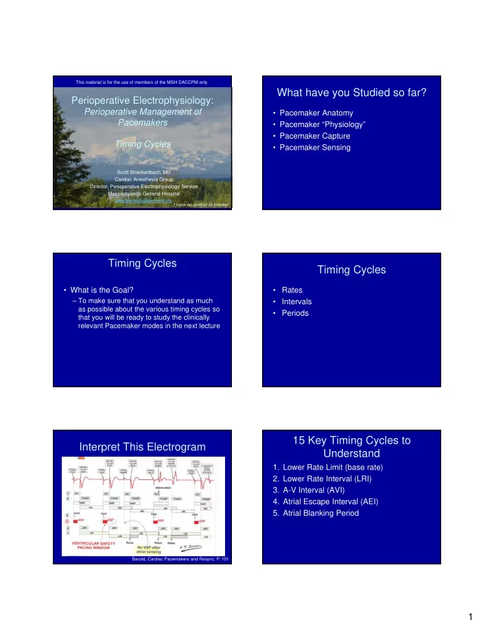

This material is for the use of members of the MGH DACCPM only What have you Studied so far? Perioperative Electrophysiology: Perioperative Management of • Pacemaker Anatomy Pacemakers • Pacemaker “Physiology” • Pacemaker Capture Timing Cycles • Pacemaker Sensing Scott Streckenbach, MD Cardiac Anesthesia Group Director, Perioperative Electrophysiology Service Massachusetts General Hospital sstreckenbach@partners.org I have no conflict of Interest Timing Cycles Timing Cycles • What is the Goal? • Rates – To make sure that you understand as much • Intervals as possible about the various timing cycles so • Periods that you will be ready to study the clinically relevant Pacemaker modes in the next lecture 15 Key Timing Cycles to Interpret This Electrogram Understand 1. Lower Rate Limit (base rate) 2. Lower Rate Interval (LRI) 3. A-V Interval (AVI) 4. Atrial Escape Interval (AEI) 5. Atrial Blanking Period Barold, Cardiac Pacemakers and Resync. P. 101 1
15 Key Timing Cycles to 15 Key Timing Cycles to Understand Understand 6. Atrial Refractory Period (ARP) 11.Post-Atrial Ventricular Blanking Period (PAVB) 7. Ventricular Blanking Period (VB) 12.Crosstalk Detection Window (CDW) 8. Ventricular Refractory Period (VRP) 13.Total Atrial Refractory Period (TARP) 9. Post-Ventricular Atrial Blanking Period (PVAB) 14.Upper Rate Interval (URI) 10.Post-Ventricular Atrial Refractory Period 15.Maximum Tracking Rate (MTR) (PVARP) 1. Lower Rate Limit Lower Rate Limit Example • The base pacing rate: – Asynchronous pacers—always pace at this rate – Demand pacers—pace at this rate if intrinsic rhythm is below the base rate • Described in beats per minute – 60 beats per minute Lower Rate Limit Example 2. Lower Rate Interval • The time between one sensed or paced event and the next paced event • Determined by the programmed lower rate limit • Described in msec 2
How does one convert Rate to Rate to Interval Conversion an Interval? • Rates are described as beats per minute Step 1: Convert Rate to beats/msec • Intervals are described as msec per beat beats X 1 min X 1 sec Rate = = beats/60,000 msec min 60 secs 1000 msec Example: Assume rate = 60 bpm = 60 beats/60,000 msec = 1 beat/1000 msec How does one convert Rate to Key Concept an Interval? Step 2: Take the reciprocal of the rate RATE INTERVAL The interval is the inverse 1 beat/1000 msec 1000 msec/beat of the rate Rate to Interval Calculation Interval Calculation • Assume the pacemaker’s rate is set at 75 bpm. How much time should elapse between one beat and the next? In other words, what is the interval? Moses, A Practical Guide to Pacing Appendix II 3
Example Example • If rate=75 bpm: • If the Heart Rate at which VF is detected is 180, what is the R-R interval at which VF is detected? Interval = 60,000 msec 75 beats • Interval = 60,000 msec/180 beats = 333 msec Interval = 800 msec • Thus an R-R interval of 333 is bad! Take Home Message • Normal rhythms have longer intervals This conversion chart lists the interval associated with 60 bpm 1000 msec paced rates from 30 to 300 75 bpm 800 msec 100 bpm 600 msec • Arrhythmias have shorter intervals 150 bpm 400 msec 200 bpm 300 msec Moses, A Practical Guide to Pacing, p. 204 Interval to Rate Conversion Interval to Rate Conversion • What if you know the interval between two • If the Lower Rate Interval is 800 msec, paced beats and you want to determine what is the Lower Rate in beats per what the paced rate is? minute? Rate = 60,000 msec/min 800 msec/beat = 75 beats/min Moses, A Practical Guide to Pacing Appendix II 4
“Programmed” vs “Derived” Interval Abbreviations Intervals • Programmed Intervals • “P” Intrinsic atrial depolarization – Lower Rate Limit (LRL) interval • “R” Intrinsic vent. depolarization – AV Interval (AVI) • “A” Atrial paced event • Derived Intervals • “V” Ventricular paced event – Atrial Escape Interval (AEI) Interval Examples 2. Lower Rate Interval • A-R: A-pace, spontaneous QRS • The time in msec between one sensed or paced event and the next paced event • P-V Spontaneous P followed by V-pace • Reciprocal of the programmed lower rate • A-V A-V paced limit • P-R Spontaneous P-QRS 2. Lower Rate Interval Example 2. Lower Rate Interval Example 1000 msec 1000 msec • The pacing mode is VVI—the LRL represents the lower • Three separate intervals: rate interval. 1. VP-VP—full 1000 msec – If lower rate set at 60, the LR interval will be 1000 msec 2. VP-VS—less than 1000 msec 3. VS-VP—slightly less than 1000 msec Ellenbogen, Clinical Cardiac Pacing, Defib, and Resync. 4 th ed Ellenbogen, Clinical Cardiac Pacing, Defib, and Resync. 4 th ed 5
2. Lower Rate Interval Example Automatic Interval 1000 msec • The time between two paced beats when the pacer is pacing at the lower rate limit Automatic Escape • If LRL is 60, the Automatic interval is 1000 msec • Three separate intervals: 1. VP-VP—full 1000 msec 2. VP-VS—less than 1000 msec 3. VS-VP—slightly less than 1000 msec Barold, Cardiac Pacemakers and Resynch. Ellenbogen, Clinical Cardiac Pacing, Defib, and Resync. 4 th ed Escape Interval 3. AV Interval • Escape Interval—the period, measured in • The interval between an atrial event milliseconds, between a sensed cardiac event (sensed or paced) and the paced and the next pacemaker output pulse ventricular event • Represents the P-R interval • A programmed interval • Usually 160-240 msec Barold, Cardiac Pacemakers and Resynch. 3. AV Interval Why are there 2 AVIs? • With both AVIs, we want the functional atrial kick to occur a given # of msec before the VP event • The pAVI starts as soon as the AP event occurs • The sAVI starts later, not until the atrial depolarization is already moving into the atrial tissue where the lead is • To ensure that the same amount of time elapses between the functional atrial kick, the sAVI is set shorter by approx 30-50 sec Barold, Cardiac Pacemakers and Resynch. 6
Paced AVI vs Sensed AVI Two AV Intervals • The Paced AV interval (pAVI) will usually be programmed approximately 30-50 msec longer than a Sensed AV interval (sAVI) – This compensates for the fact that the pAVI timing circuit (stopwatch) starts as soon as the atrium is paced—this happens 30-50 msec before the atrial depolarization is sensed by the atrial pacing electrodes Paced AV Delay vs Sensed AV Medtronic Temp Pacer 5392 Delay What is Rate Responsive AV Delay? Question? How long after an intrinsic P-wave will the pacer wait for a spontaneous R-wave (QRS) before firing a V-pacing spike? Answer=140 msec 7
4. Atrial Escape Interval 4. Atrial Escape Interval • Atrial Escape Interval (AEI)—the period in a dual chamber pacemaker’s timing cycle Pacing interval (LRL) initiated by a ventricular sensed or paced Atrial Escape Interval AVI event and ending with the next atrial paced event. • This is a derived interval – Depends on the programmed LRL and the AVI 4. Atrial Escape Interval AEI=VAI Atrial Escape Interval (AEI) often called the Ventricular-Atrial Interval (VAI) What would happen if a P wave occurred at the arrow? What would happen if a PVC occurred at the arrow? Barold, Cardiac Pacemakers and Resynch. p.92 Ventricular-Atrial Interval With the LRL, LRI, AVI, and AEI you can program any VOO, AOO or DOO pacemaker 8
Sensing Revisited Pacer sensors depend on signal amplitude If you want to use the sensing and slew rate to detect appropriate signals function of the pacer, many more such as the P-wave or R-wave. timing cycles are needed The Sensors also need methods to avoid What kinds of Signals can be detection of inappropriate signals that can negatively affect the pacer function sensed inappropriately? Detectable Signals after a 1. Same Chamber Signals Paced Beat • Same chamber signals • Stimulus artifact • Far-field signals • After-depolarization • Evoked Potential (QRS/P-wave) • Repolarization (T-wave) 9
Detectable Signals on Detectable Signals on the Atrial Ventricular Channel Channel • Atrial paced stimulus • Atrial paced stimulus after-potential • Evoked response (P-wave) • Spontaneous P-wave Barold, Cardiac Pacemakers and Resynch. p.64 2. Far-Field Signals Far-Field Signals: VA Crosstalk • Atrial pacing artifacts sensed on V-channel – AV Crosstalk • Vent pacing artifacts or QRS sensed on A- channel – VA Crosstalk Ventricular pacing stimulus and evoked QRS are sensed on the AEGM Barold, Cardiac Pacemakers and Resynch. Sensing-Related Timing Cycles How does the Pacemaker • Blanking Periods minimize the likelihood of • Refractory Periods • Cross Talk Periods Sensing Inappropriate Signals? 10
Recommend
More recommend