SLIDE 1
1
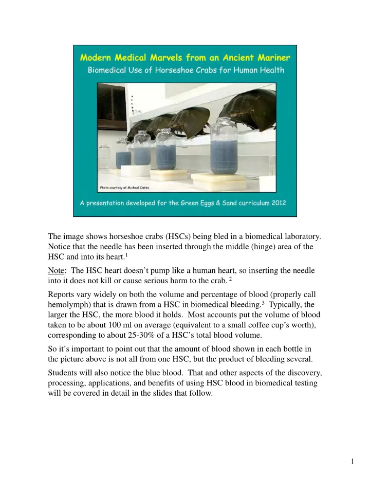
The image shows horseshoe crabs (HSCs) being bled in a biomedical laboratory. Notice that the needle has been inserted through the middle (hinge) area of the HSC and into its heart.1 Note: The HSC heart doesn’t pump like a human heart, so inserting the needle into it does not kill or cause serious harm to the crab. 2 Reports vary widely on both the volume and percentage of blood (properly call hemolymph) that is drawn from a HSC in biomedical bleeding.3 Typically, the larger the HSC, the more blood it holds. Most accounts put the volume of blood taken to be about 100 ml on average (equivalent to a small coffee cup’s worth), corresponding to about 25-30% of a HSC’s total blood volume. So it’s important to point out that the amount of blood shown in each bottle in the picture above is not all from one HSC, but the product of bleeding several. Students will also notice the blue blood. That and other aspects of the discovery, processing, applications, and benefits of using HSC blood in biomedical testing will be covered in detail in the slides that follow.
SLIDE 2
2
You can almost guarantee that anyone watching this slide show has benefited in some way, or will benefit at some time from, the biomedical use of horseshoe crabs. Some may know that this has something to do with HSC blood, and a few may have heard that this involves making sure vaccines are safe to use, but most won’t know about these other materials that also need to be tested with it. In addition to the above-mentioned, required-by-the-FDA examples of medical materials that are tested with LAL, there are several other instances where LAL may be used to screen or assess other medicines for potential health threats to humans. An example of this is testing contact lenses or contact lens solutions that are suspect in causing bacterial eye infections, such as keratitis, an inflammation of the cornea4 (which may also be caused by fungal infections or the Herpes virus).
SLIDE 3
3
In addition to every man, woman and child near and dear to us, the health and well-being of our pets and many other domesticated animals also benefits from this test! As with medicines used in humans, all veterinary injectables and implantables in the U.S. are tested with HSC blood product to ensure that they are safe to use. The reasons for this are the same as for humans – contamination of any veterinary medicines that come into direct contact with blood and tissue can cause the same kind of fever and illness reactions in dogs, cats, horses, etc. as they do in humans.
SLIDE 4
If you’re wrapping this ppt around the LAL saliva test, this is a good place to set up the experiment. Explain to students that you are going to perform an experiment that simulates the use of LAL (the product derived from HSC blood) in biomedical testing. Hold up one of the vials and explain that these are real vials of the same stuff (containing the white powder derived from HSC blood) that is used biomedically to test vaccines and other medicines used in humans. We will use these vials to do a simple demonstration of the gel-clot reaction, using one vial to test for the presence of endotoxins in a source readily available to us – our mouth – and the other as a control to test for endotoxins in a sample of purified (bottled) water. If students will also be doing these tests at their seats, refer them to the LAL lab demo instructions handout provided for that purpose. One key point about this – when you direct students to swill some of the pure water around their mouths (to get the saliva sample), emphasize just taking a little sip of the water (too big a sip may dilute the endotoxins too much & cause a negative test). We have found that the biggest challenge for students in doing the set-up is with their pipetting skills, so if possible build in some time to have students practice and master those skills with samples of the purified water before they actually try to dispense their samples into the vials. Caution students not to grasp the pipettes by the stem end, but by the bulb (so they don’t introduce a source of contamination to the sample). Also emphasize and demonstrate the process of dispensing and/or holding steady the level of liquid in the pipette. If air bubbles are introduced (easy to do with the saliva), have them start over. Also don’t forget to have students gently swirl each of the vials (to ensure mixing of the sample with the LAL powder) before setting them aside to incubate. Once all the vials have been set-up, direct students to set them aside and leave them undisturbed for a period of time (15-30 minutes is typically good) before observing the results of the test.
4
SLIDE 5 5
The slides to follow will give some of the history of why and how this test came to be. Up to the mid-1800’s, diseases were thought to be caused by spontaneous generation, excess ‘humors’, or even demons, (the latter as punishment for a person’s misdeeds). Eventually, thanks to the work of Louis Pasteur and others, the germ theory of disease, or the idea that microbes were causal agents for certain diseases, came to be accepted. In time, use of the microscope allowed for the association of various forms of bacteria with particular diseases, such as anthrax, smallpox, typhoid fever, the plague, etc. The first recorded cases of doctors injecting substances into their patients came out of Europe in the mid-1600’s – including such things as ale, opium, wine, urine, and various putrid waste products. The development by Edward Jenner (1796) of a smallpox vaccine, made by taking the pus from the cowpox sores of a milkmaid and injecting it into an 8-year
- ld boy (to confer immunity to smallpox) is an interesting side story to all this. 5
In the 1800’s, with the development and use of more vaccines, injections became more
- common. But doctors began noticing side reactions to these injections, including
inflammation around the shot site, often followed by malaise,* fever, low blood pressure, shock, and even death. Now if you’re a physician – providing a treatment to make a sick person well or a vaccine to keep a well person from getting sick – then fever, malaise, shock, and death, would hardly be desired outcomes of your efforts! * malaise - a vague feeling of discomfort, often at the onset of an illness; can involve a feeling of exhaustion, or of not having enough energy to accomplish usual activities. (from the French "mal" (bad or ill) + "aise" (ease) = ill at ease).
SLIDE 6
6
For some time, the actual source or cause of injection fever remained a great mystery. Lacking a better explanation, the cause of injection fever was attributed to the body’s response at being pricked by a needle. In the meantime - as scientists sometimes do, when they don’t have a clear answer for something - they concocted a fancy-sounding name for this mystery source. They called the unknown agents of injection fever ‘PYROGENS’ - from the Greek Pyros (for ‘fire’).
SLIDE 7
7 There’s a fascinating history of scientific discovery that led to identifying the actual source of these pyrogens, including the work of Lister (1861), Koch (1880), Pfeiffer (1892), Hort & Penfold (1912) and Seibert (1923). As a result of their efforts, the cause of injection fever was finally traced to the lipopolysaccharides (LPS) from the cell membrane of gram-negative bacteria. Gram-negative bacteria (GNB) have been described as “thin-skinned”. Just as we humans routinely shed outer layers of skin, bits of GNB-LPS layer are sloughed off as they move. When GNB are killed, even larger bits of LPS/endotoxin are released. When these endotoxins appear in the blood, our immune system detects them, and one response is to raise our body temperature (as a way of killing off the infection). Normally, in small doses, this is not a problem, but at higher levels and higher temperatures, fever is induced, and if intense or prolonged, this can be deadly. 6 Because these toxic materials are derived from structural components of the bacterial cell (not substances that were produced by, and released externally from, the cell), scientists came up with another fancy name for them. They called them endotoxins. For an in-depth immersion in the significance of the LPS layer of gram-negative bacteria as an activator of various critical immune system pathways in animals ranging from HSCs to humans, check out the article by Alexander and Rietschel (2001).7
SLIDE 8 8 This chart summarizes the two main groups of bacteria that came to be recognized and some key differences between the two. Although both groups include some nasty characters in terms of human disease, the gram-negatives are especially pervasive and problematic. The gram-positive cell wall is thick and made up of interlocked (mesh-like) layers
- f a molecule called peptidoglycan. Since gram-positive bacteria are largely
terrestrial, this thick cell wall gives them a rigid shape and keeps them from drying
- ut in hostile environments.6
Gram-negative bacteria have a thinner cell membrane. It features a single inner layer of peptidoglycan (for strength) and a thicker outer layer of lipopolysaccharide. Gram-negative bacteria are found mainly in aquatic environments, where their thinner, less-rigid cell membrane offers more flexibility for moving in water. 6 It should be noted that there are many other sources (both biological and non- biological) of pyrogens. This includes prostaglandins from the cell wall of gram- positive bacteria and certain fungi But these substances are on the order of 50,000 times less pyrogenic than endotoxins from gram-negative bacteria. 8 That and the fact that extensive research has shown endotoxins to be the one pyrogen source most likely to contaminate injectable drugs and devices, is a big reason why the search for a way of screening such medicines for endotoxin became an essential concern for the biomedical industry. 9
SLIDE 9 9
So what’s the big deal about endotoxins? Go down the list. Gram-negative bacteria are ubiquitous in the environment. We drink them, eat them, pick them up from things we touch, and even breathe them. And often, the very act of killing bacteria releases ‘free’ endotoxins to where they can do us harm. Chemical or physical processes are ineffective in controlling them. Antibiotics don’t destroy them, nor does standard steam sterilization Endotoxins are also the most potent pyrogen known to man. So what is an ‘ng’? An ng is a nanogram = one-millionth of a milligram, or one trillionth of a kilogram. This means that it
- nly takes very minute levels of endotoxin to produce a fever reaction in humans.
Endotoxins can also cause profound inflammation of any tissue that is exposed to them, which if severe enough, can lead to impaired function of the lungs, brain, kidneys, etc. If fever is prolonged, it can lead to tissue breakdown, shock and ultimately death. One of the challenges of preparing injectable medicines with levels of endotoxins that are safe for use in humans is that of finding endotoxin-free sources of the raw material water (called “Water for Injection” or WFI) used in preparing injectable medicines. These waters are derived from various natural sources (both surface waters and ground water) that contain varying amounts of endotoxins. Distillation is the preferred method for making this WFI endotoxin-free. But problems can arise via tranfer or contamination of storage containers and delivery devices. And many gram-negative bacteria can grow and survive in distilled water at low ambient temperatures (some for well over a year); and even if the bacteria are killed off, the endotoxins remain behind to cause problems.8 Note: Some of the few cases of patients getting ill from medicines that had passed the LAL test were found to be caused by contaminated storage containers or pumps used to deliver WFI that was mixed in with (otherwise safe) meds administered in IV systems.8
SLIDE 10 10
We already know the bad news about endotoxins - if they get into our blood, they can make us sick. The good news is that it’s OK to drink them and eat them - inside our closed digestive system they are not a problem. In fact, endotoxins are present in our food and water, and are even produced by bacteria in our mouth and intestines. There are several ways that a healthy body deals with food and drink delivered endotoxins. Acids in the stomach kill many bacteria, minimizing production of further endotoxins. Then there is the physical barrier provided by the stomach lining and intestinal mucosa. And any endotoxins that manage to cross these barriers are inspected by, and typically removed by, cells and proteins of the human immune system. But the body’s main defense against endotoxins reaching the blood from the digestive system is the liver. The products of food digestion are absorbed from the small intestine by capillaries that deliver them directly to the liver via the hepatic portal vein. The liver plays the role of ‘gatekeeper’ filtering and detoxifying any digestive endotoxins before they get passed out into the blood. 10 Problems with endotoxins from foods can arise when any breakdown in any of these levels
- f protection occurs, such as stomach ulcers or ulcerative colitis (impacting the intestinal
lining), and hepatitis or cirrhosis (impairing the liver’s normal detoxifying function). 11 But what happens when we receive and injection? (click to advance the slide animation) That blood goes to the heart and circulation before it can be screened by the liver, and thus gets pumped and delivered to the cells and tissues of the brain, kidneys and various other parts of the body, where the endotoxins can cause fever and other reactions. Note: In reference to the fact that injectable medicines bypass the digestive system, they are also described as PARENTERAL - from the Greek “para” (beyond) & “enteral” (gut).
SLIDE 11 11
With the increasing use of injectable medicines in the 1900’s, and especially with the introduction of intravenous drugs in treating battlefield-wounded soldiers in WW1, the need for a test to screen medicines for endotoxins intensified. In looking for an animal model, lab mice and rats were not found to be especially
- sensitive. Dogs and horses were sensitive, but not the most desirable lab animals.
The rabbit proved best – they were easy to keep, care for, and work with, and pound per pound, they showed a very similar response to endotoxins as humans. The rabbit pyrogen test thus became the gold standard for pyrogen testing in 1942, when it was introduced into the USP (United States Pharmacopoeia).
SLIDE 12
12
In the rabbit pyrogen test, a sample of the test medicine was administered to 3 rabbits (by way of an injection of a measured quantity of the medicine into the rabbits’ ear). Rectal probes were inserted into each rabbit to monitor their temperature. Side note: Technicians performing these early rabbit tests got very good at cradling rabbit in their lap (to reduce stress) while inserting the rectal thermometer (resulted in the test also being called “the lap test”), but when new techs came on, who weren’t as good with the rabbits, this could cause anxiety in rabbits affecting the test results.12 If after 3 hours, the sum of the temperature increases observed in the 3 rabbits was less than 1.4 °C, the medicines passed, and were deemed safe for use in humans. But if the temperature increases for the 3 rabbits was equal to or greater than 1.4°C, then 5 additional rabbits had to be tested … Then, if the sum of all 8 rabbits temperature increases was equal to or greater than 3.7°C, the meds were considered unsafe for use. If the temperature increases for the 8 rabbits totaled less than 3.7°C, the meds passed and were deemed safe for use. 12 Note: failure to pass the rabbit pyrogen test (or the LAL test that replaced it) did not necessarily mean that the whole batch of medicine had to be tossed away. At that point, dilution of the medicine, either with other batches that did pass the test, or with new (untested) batches that were more rigorously prepared, could be done and the resulting combination batches retested. Such dilutions/mixings could be done until the test was passed, thus minimizing product loss.
SLIDE 13
13
But the rabbit test had its problems. Not only were conditions difficult to control – due to individual differences in the rabbits, excitability factors, and other variables – but the tests also yielded many false positives (thinking the medicine failed the test, when it was actually safe to use) and false negatives (medicines passing the test, when they weren’t safe for use). The latter results were especially troublesome, including several health care incidents in the 1970’s from meds that had passed the rabbit test: 1) In 1972, 39 cases of life-threatening aseptic meningitis occurred in patients receiving radiotracer injections for assessing the flow of cerebrospinal fluid. Cooper and Harbert found endotoxins to be the cause, and that endotoxins were at least 1000x as toxic when administered intrathecally (into the sheath surrounding the spinal cord) as compared to typical bloodstream pathways. They concluded that the rabbit test was not sensitive enough to pick up these lower endotoxin levels (but the LAL test was). 12 2) In 1974, an outbreak of endotoxin-induced fever-reactions occurred in patients receiving rabbit-test-approved serum albumin for trauma and severe disease therapies. Such reactions were subsequently minimized by using LAL to test albumin supplies.8 3) In 1976, after receiving swine flu vaccine, 30 people died and several hundred people developed Guillain-Barré syndrome 13 (a rare condition in which the body’s immune system attacks the nerves causing temporary muscle weakness or paralysis). Later testing with LAL indicated high levels of endotoxins in the vaccine that (had this been known at the time) might have prevented use of these vaccines, and the adverse health problems that resulted. 9
SLIDE 14 14
Such problems prompted health care officials to seek out an alternative, more reliable way of screening for bacterial endotoxins. Enter Limulus and Doctors Fred Bang & Jack Levin! As with many important scientific discoveries, this involved some serendipity, along with a measure of that old “chance favors the prepared mind” adage. Back in 1950, Dr. Frederick Bang, a researcher at Woods Hole Oceanographic Institute on Cape Cod, was investigating marine animal immune systems. Due to its ancient lineage, he considered the HSC a good candidate for revealing primitive immunological functions. He started injecting HSC blood with various kinds of bacteria, and hit the jackpot with one called Vibrio. Here is a quote from Bang’s (1955) ground-breaking discovery: 14 “Bacteria obtained at random from fresh seawater were injected into a series of horseshoe crabs (L. polyphemus) of varying sizes. One Limulus became sluggish and apparently ill. Blood from its heart did not clot when drawn and placed on glass, and yet instant clotting is a characteristic of normal Limulus blood … Other gram-negative bacteria or toxins also provoked intravascular clotting in normal Limuli. When these same bacteria or toxins were added to sera from normal Limuli, a stable gel was formed.” Upon further testing, Bang was able to show that heat-killed extracts of these bacteria also induced clotting. Although these were not the kind of results he expected to find, like all good scientists, Dr. Bang was smart enough to see that he was on to something significant. He was also wise enough to see that he could use some collaboration, so he hooked up with
- Dr. Jack Levin, a specialist in hematology from Johns Hopkins, and together they started to
unravel the mystery that ultimately led to the development of the LAL test.
SLIDE 15 15
But these discoveries hardly happened overnight either. Many year of research and levels of experimentation ensued. In one of the experiments, Bang and Levin separated out the HSC blood cells (amebocytes) from the plasma (hemolymph). They found that plasma without cells would not clot in the presence of endotoxins. This clued them that the clotting factors were derived from within the amebocytes (the only kind of blood cell HSCs have). 12 Eventually, they were able to trace those clotting factors to proteins found in granules within the amebocytes. When exposed to harmful bacteria, these amebocytes change shape (developing long pseudopod-like processes), and the granules “pop out’ or are released out of the cell. Further investigation revealed that proteins released from the granules triggered a cascade of enzymatic reactions that caused the blood of the HSC to form a clot that engulfs and immobilizes the bacteria and/or
- endotoxins. Subsequent research enabled them to isolate and refine this
biochemical process and duplicate it inside a test tube. Development of the LAL test was the end product of Bang and Levin’s outstanding research and collaboration. 12
SLIDE 16 16
Even though LAL was shown to offer a much more reliable and sensitive tool for endotoxin screening, it took some time for the test to become accepted, used and approved as the standard for pharmaceutical testing. Although developed by the 1960’s, and in use by the medical community by the early 1970’s, it wasn’t until 1977 that the FDA officially approved its use (as an alternative to the rabbit test) for testing injectable medicines and until 1979 that it was approved for testing implantable medical devices. There were several reasons for the delay. The reluctance of pharmaceutical companies to make a major shift in testing procedures was one issue. Highly variable results with early LAL tests didn’t help - though this was later attributed to bleeding of juvenile HSCs (which have fewer amebocytes than adults). 2 and was subsequently corrected. A big breakthrough occurred in 1990, when a conference of the U.S. Pharmacopeia (USP) aimed at minimizing use of animals in pharmaceutical testing, removed the rabbit test from the USP, making the LAL test the only viable tool for endotoxin screening. Although a major improvement over the rabbit test, LAL did not solve all problems and is not considered foolproof. One of the major drawbacks of LAL is that it only detects levels
- f endotoxins. It cannot identify which kind of bacteria releases the endotoxins. This
greatly limits its value as a diagnostic tool. Also contaminations of drugs with gram- positive bacteria (e.g. Staphylococcus species or Bacillus cereus, common contaminants in pharmaceutical manufacturing), are not an unlikely event. 8 Cases of patients getting ill from use of human growth hormone and other LAL-test approved animal serums have also been noted. 8 Later studies indicated interference factors from LPS-neutralizing and degrading blood processes that limited the gel-clot reaction.
SLIDE 17
17 So, why do HSCs have this extraordinary sensitivity? If you think about it, HSCs have been living in a virtual seawater soup for hundreds of millions of years, exposed to a myriad of microbes that could infect them, make them sick and even kill them. A single teaspoon of seawater may contain billions of gram-negative bacteria! So how do they keep the harmful microbes at bay? Like all invertebrates, HSCs are cold-blooded/ectotherms (so they can’t use fever to heat up and kill off bacteria like we do), and they lack antibodies (to detect, recognize and confer immunity to foreign invaders). HSCs also have an open circulatory system, meaning that harmful bacteria, upon entry into their bloodstream, can quickly gain access to many of the HSCs’ internal organs and tissues. In contrast to humans, HSCs also are limited to just one kind of blood cell (called an amebocyte) in their immune system defense arsenal.16 So how indeed do they do it? Despite its apparent simplicity, over those millions of years of exposure to all sorts of microbes, the HSC has evolved a highly efficient and sensitive system for detecting and dealing with such invaders. So when an HSC is wounded (as in the picture above), the amebocytes react quickly by releasing enzymes from their granules (found in the cytoplasm) into the blood around the wound site. This triggers the gel-clot reaction, which not only seals off the wound site from further invasion, but also acts to neutralize the bacteria and endotoxins present, thus buying time for other immune system weapons (such as peptides and other proteins found in the blood) to activate and destroy them. 17
SLIDE 18 18
- Dr. Ronald Berzofsky - the biomedical scientist featured in this video –
has been involved in applications of LAL for decades, including ground- breaking research in developing recombinant Factor C, a genetically- engineered form of LAL that has potential to negate the future need for bleeding HSCs to produce LAL test media. 18
- Dr. Berzofsky has also been extremely helpful in bringing the biomedical
side of the story to GE&S - both in presenting at our Delaware workshops, and in providing us with much of the material and perspectives used in development of this powerpoint. He has also played a key role in guiding initial development, and ongoing improvement of the LAL-Lab activity, as well as supplying us with thousands of free vials of LAL media that we provide to teachers for doing gel-clot tests with their classes. This clip features Ron expounding – in his own inimitable way – on the basics of what is going on with the HSC gel-clot reaction.
SLIDE 19 19 This slide in offered as a point of reference. Go over the list row-by-row. Note: some
- f the information included in the notes section of slide 16 could apply here as well.
Although complex, antibody-based, immune systems are only found in vertebrates, this doesn’t mean that the immune systems of invertebrates are poor or ineffective. As we’ve seen with the HSC, these animals have evolved other weapons and strategies! Anyone who sees the HSC as primitive, should read the chapter on the HSC immune system in The American Horseshoe Crab (Shuster, Barlow, and Brockmann, 2003). 17 Also this is as good a place as any to demystify the blue blood part of the HSC story. Firstly, the blood of the HSC inside the animal is actually more of a straw color. Only when the blood is exposed to air does the copper pigment in it cause it to oxidize blue. And the blue color has nothing to do with endotoxin-detecting capacities of its blood. HSCs are also not the only blue bloods. Molluscs (clams, snails, squid and octopi), crustaceans ( lobsters, shrimp, blue crabs, etc.) and scorpions also have blue-blood. Questions often arise as to why the blood in human veins appears blue under our skin. The answer has to do with how the light spectrum is reflected or refracted by our skin. Veins appear blue because only the high-energy, low-frequency blue wavelengths of light are reflected by the veins, while the other colors are absorbed by these vessels. In reality, venous blood tends to be a dark, dirty red because it is unoxidized and carries
- wastes. Arterial blood, having been freshly oxygenated in the lungs, appears bright red,
because its iron-based pigment, hemoglobin, oxidizes red like rust.
SLIDE 20
20
Due to the design of the HSC circulatory system, bleeding only removes blood from its large pericardial sinus (heart) and adjacent large arteries. Most of the remaining hemolymph (roughly 70% of the total blood volume) remains in the spongy tissues of the prosoma and diffuses slowly into the large vessels.12 One may notice in these and other video clips or images of HSCs being bled that it is done in a sterile lab environment with technicians wearing masks, lab coats and other protective equipment. Might be interesting to ask students why they think this is the case. Is it to protect the humans or protect the crabs? Actually it is neither. It’s all about protecting the blood product that is to be used in making the LAL. Why would this be important? If conditions were not sterile, blood drawn from the HSC would be exposed to bacteria/endotoxins, causing amebocytes to release clotting proteins into the blood & induce a clot. Think about it! If the blood is already clotted before or while it is being turned into a product that relies on a clotting reaction to be useful as a test - well that just wouldn’t work out, would it? That’s why the bleeding process takes place in sterile chambers where ambient air, water, surfaces, etc., as well as the humans entering or leaving them, are carefully monitored and controlled.
SLIDE 21
21 In China - where crabs are also bled for biomedical use - the product is called TAL, for Tachypleus Amebocyte Lysate and in India, where the Indian HSC is used, it is referred to as CAL for Carcinoscorpius Amebocyte Lysate. The more generic term “Lysate” is also often used to describe any of these products. In recent years, realization of the economic potential of biomedical use of HSCs has prompted concerns about potential overexploitation in parts of the world (e.g. India and Southeast Asia) where HSC populations are not nearly as robust and regulated as in North America, where production of LAL is a multi-million dollar industry. 19 One of the questions teachers often ask is: “How many vials of LAL are produced from the bleeding of one HSC?” Another is: “How much is that product worth?” Thanks to information provided by Dr. Ron Berzofsky (personal communication),20 we took a stab at answering those questions, as follows: Firstly, we assume an average volume of 100 ml of blood product per HSC bleeding. Then, according to Dr. Berzofsky, 100 ml of HSC blood, upon centrifuging, yields 5 ml of packed amebocytes, and this amount yields 425 LAL single (gel-clot) test vials (in case your wondering, each single test vial uses 0.1 mg of LAL material). Now to the economics. In 2010, single test LAL gel-clot vials market commercially in the U.S. for $4.44 each; 21 so (doing the math), the total value of LAL product derived from the bleeding of one HSC would translate to: 425 x $4.44 = $1887.
SLIDE 22 22
This is a good place to have students pause and observe the results of their LAL tests. The process is quick, easy and straightforward and well explained in the student handout (and slide above). In most cases, students will observe a positive gel-clot test in the saliva sample and a negative in the purified water control. However, if the samples are left too long before observing, sometimes the gel will have developed and start dissolving in the saliva sample, yielding a false negative
- result. That is why in real-world testing of vaccines and other medicines, a rigorous time and
temperature controlled protocol is followed to ensure that those variables do not impact the results. And then there is the statistical nature of the sampling protocol. A minimum of 3 units of a batch of medicines are tested – one near the beginning of the batch, one from the middle and one from the end of the product lot. Results from the 3 samples can be tracked individually or pooled. If done individually, a positive test from any of the 3 samples requires that repeat testing be done. If the units are pooled and test positively for endotoxins, repeat testing must be performed as follows: For the 1st repeat: twice the initial number of replicates must be run, if that fails (if the pooled samples produce a positive test for endotoxins), a 2nd repeat test is required, in which an additional 10 units are tested individually. If all 10 units pass, the meds are safe to use, but if any one of the units fails, then that batch of meds cannot be used. 8 The gel-clot is the simplest and most widely used version of the LAL test. Although the method tends to be qualitative (yes/no, pass/fail), in actual pharmaceutical lab testing, controls of known amounts of endotoxin for comparison can be used to quantify amounts of endotoxin present. It is important to emphasize that a negative LAL test doesn’t mean that there are no endotoxins, just that the levels are low enough to be deemed safe by FDA standards.
SLIDE 23
23 Nearly all of the 400,000 or so HSCs that are collected annually in the U.S. for biomedical bleeding are returned to the environment where they were harvested within 24-48 hours after collection. An exception to this occurs in Massachusetts, where regulations allow that animals bled for biomedical purposes can be passed on to commercial fishermen for use as bait in catching conch and eel. 22 The rationale behind this is that if a certain number of crabs are going to be harvested and used for bait anyway, they might as well use the biomedically-bled crabs twice (getting dual use out of one crab), as opposed to exposing still more crabs to human use. The other important thing to know about biomedical use is that the large majority of crabs do survive the bleeding process. Variation in reported mortality rates (2-30%) from bleeding are attributed in large part to how they were collected, and to a lesser extent, on conditions under which they are kept and bled. Crabs collected by dredge or trawl experience higher mortality rates than those collected by hand harvesting.3 The level of stress horseshoe crabs are exposed to during handling also factors in. One study found that crabs bled under low stress conditions, had 0% mortality, compared to 8.3% dying when exposed to high-stress handling conditions. A more recent study in Massachusetts found female HSC mortality from bleeding to be as high as 30%, much higher than had previously attributed to biomedical use. 23 Biomedical companies are constantly looking for ways to reduce negative impacts on the HSCs they use and depend on for the life-saving products they provide.
SLIDE 24 The top photo features a bioreactor – a device in which genetically-engineered products (such as Factor C) are produced, usually by growing them in bacteria. Factor C is already available, but will take some time to replace LAL – due to it being so well entrenched in pharmaceutical protocol and confirmed reliability. 18 The same challenges will face whatever comes of this latest promising alternative
- ffered up by researchers on the African clawed frog. Like the HSCs, these frogs
have also evolved a strong antibacterial defense system. In the wild, the African clawed frog produces antibacterial peptides - small chains of amino acids - on its skin to protect it from infection. Princeton researchers have found a way to attach these peptides, which can be synthesized in the lab, to a small electronic chip that emits an electrical signal when exposed to harmful bacteria, including pathogenic
- E. coli and salmonella. 24
The African frogs, which are common in labs and pet stores, are not harmed in the process, and the peptides can be synthesized. McAlpine and Manu Mannoor (pictured above, the latter a Princeton graduate student who worked on the project), hope that technology based on their electronic chip will eventually replace LAL as the standard for contamination testing, negating the need for using HSC blood.
24
SLIDE 25 25
So how sensitive is LAL to endotoxin? Call on students to read the two blurbs on the slide (credit John Dubczak, of Charles River Laboratories, for the swimming pool analogy). It’s also been noted that the the LAL test is so sensitive that it can pick up the tiny bit of endotoxin produced from a single E. coli bacterium in a ml of water! If time allows, this could be a good place to mention the more quantitatively precise variations of the LAL test that have been developed. This includes: 12,25 1) Turbidimetric systems measure (using a spectrophotometer) the level of cloudiness or turbidity in the coagulogen that is produced by the gel clot reaction after incubation at a fixed temperature over a fixed time period (the higher the turbidity, the greater the concentration of endotoxins). 2) Chromogenic assays utilize a chromogen, or chemical that changes color in response to the amount of endotoxin (again at fixed temp. and time period). The higher the endotoxin concentration, the more chromagen that is released (as measured by an
- ptical reading device tuned to chromagen wavelength).
3) Kinetic assays measure the rate of change in turbidity or color during the assay (based
- n the premise that the higher the concentration of endotoxin, the more rapidly the
reaction being measured will take place). Kinetic assays can provide a greater sensitivity over a wider range than turbidimetric or chromagenic endpoint assays, but require more elaborate instrumentation. 4) Portable, hand-held electronic versions of the LAL test are now available that allow for simplified, fast (15-minute) and accurate testing in the lab or field.
SLIDE 26
26
This slide brings us back full-circle to where we started. These are the reasons why we all depend so much on this test derived from the HSC Students may wonder how things like needles, bags, catheters, valves, pins, plates, and other solid medical devices can be tested with LAL (since the gel-clot test utilizes a liquid material). Generally these devices are rinsed with endotoxin-free water and then the rinsate is tested. In some cases, there are protocols by which the medical device itself is immersed in a certain quantity of LAL reagent and the reaction of endotoxins that tightly adhere to the surface of that device is measured. The chromogenic test is usually used so that it can be quantitatively scored. 9 Outside of the human health field, the sensitivity of LAL to bacterial levels has resulted in its use for various environmental and even industrial applications. This includes certain applications for the monitoring of water and air quality. Endotoxins in the air can cause damage to human lungs; cases of respiratory problems linked to high levels of endotoxins have been reported from people breathing the air around sewage treatment plants, cotton milling plants and even from concentrated cigarette smoke. 9 LAL testing for endotoxins is also common in the food industry, where it is used to ensure a higher purity of meats, fish, milk and other products destined for human or animal consumption.9
SLIDE 27
27 So – bottom line – what does all of this mean to you and me? The answer is “a lot!” In addition to ensuring that our vaccines, insulin, allergy shots, IV’s, and other injectable and implantable meds are free from contaminants that could make us sick, LAL has also greatly improved the speed of diagnosis and treatment for certain bacterial diseases, including spinal meningitis and some urinary tract infections. 9 Bacterial sepsis - a condition that kills thousands of hospital patients every year - also has potential to be greatly reduced through use of LAL for rapid diagnosis. As with diagnosing meningitis, LAL can’t identify the kind of bacteria causing sepsis, just that it’s bacterial (not viral), so antibiotic treatment can be promptly administered. LAL has also been used to screen anti-endotoxin compounds for potential life-saving sepsis therapies. Interestingly enough, one of the compounds showing therapeutic potential was isolated from Limulus hemolymph! 9 And many other specialized applications are also in use. Reference picture above: In the LAL video segment accompanying this lesson, there’s an interesting piece at the end on using LAL to ensure that lab-cultured skin tissue for burn victims is safe. There’s a rather tight window of time for use of these tissue-cultured skin samples. Before LAL, they would have had to use a petri dish approach to test for bacterial contamination, taking at least 24 hours. By using LAL, tissues can be screened in an hour, greatly speeding up the healing and recovery prospects for burn patients. Another special application relates to artificial kidneys and the water used to prime dialysis machines, both of which can be contaminated in use/reuse with endotoxins. Adverse reactions in artificial kidney/dialysis patients have been noted where use of LAL in monitoring the equipment has not been as rigorous as it should be. 9
SLIDE 28 28 If it’s not already doing enough for us on Earth, LAL is now being used in outer space! NASA , with help from Charles River Laboratories, has developed a mini-LAL-lab called LOCAD-PTS (Lab-On-a-Chip Application Development–Portable Test System) for use on the International Space Station, the space shuttle and other space missions. Thanks to clever engineering and the rapid sensitivity of HSC enzymes, this device is small, sleek, and fast (not unlike the ‘tricorder’ device of recent Star Trek films!). 26 Use in space involves swabbing the spacecraft instrument panels and other surfaces, and inserting the swab contents into one of several narrow channels in the LOCAD-PTS. Each channel contains dried LAL powder and a colorless liquid, which turns green in the presence of bacteria and fungi (the darker the green, the greater the contamination). Results are available in 15 minutes - much faster than the old (3-day) petri dish culture
- method. Astronaut health is similarly monitored with this device, requiring only tiny
amounts of body fluids to be tested for bacterial infection. In a recent space station mission, Astronaut Sunita Williams (note HSC earrings above) tested this technology. 26 NASA also plans to use this technology to analyze samples from spacecraft surfaces before launch to verify that they are free of microbes and other organic material. The rigorous cleaning process is required by the Planetary Protection Act, established by NASA to ensure spacecraft don't contaminate pristine environments on other planets with Earth microbes. And given the incredible sensitivity of LAL to microbial life, NASA is also exploring whether the PTS gizmo could be sent into space for use in testing soils scooped up by probes from other planets for the presence of microbial life.
SLIDE 29 29 Given the wonders of LAL, researchers are looking to the HSC for a wealth of other biomedical applications, and they are finding them! HSC blood has also proven sensitive to glucans, a component of the fungal cell wall, resulting in a variation of LAL (called the Fungal G-test) that is now being used as a diagnostic tool for fungal infections in humans. This test has been found especially useful in early detection of Candida (yeast) and Aspergillus (toxic mold) infections, 9 both of which are widespread and can cause a wide array of human health problems. Meanwhile, researchers from India have been exploring amazing medical applications arising from the embryonic peri-vitteline fluid inside the HSC egg. They found it to be a viable medium for promoting and prolonging stem cell culture, with potential human health applications to heart, pancreas and other organ/tissue regeneration therapies. 27 In one avenue of research, researchers found that the molecule lectin, from HSC peri- vitelline fluid, had a dramatic influence on cardiac development in chicks and mice, resulting in an increase in number of cells and size of the heart. Similar applications, through use of HSC peptides to induce naïve stem cells to take a particular develop- mental pathway, may yield repair/regeneration therapies for other organs and tissues. 28 Biomedical benefits of HSC peri-vitteline fluid have also been forecast relative to its
- bserved anti-angiogenic activities. This includes potential applications to treatment of
cardiac disorders and cancer therapies, using inhibition of angiogenesis as a means of “suffocating” tumors, by depriving them of vascular irrigation and oxygen supply. 27
SLIDE 30 30
Meanwhile, researchers are finding other potential “wonder” medicines from the blood of the ancient mariner. This includes several classes of peptides (names like tachystatins, tachyplesins, and polyphemusins) that have demonstrated a capacity for killing a wide array of gram+ and gram- bacteria. Researchers at the National University of Singapore have recently synthesized HSC-derived peptides (called Sushi peptides) that show especially promising therapeutic
- potential. 29 Study of these compounds is improving understanding of how antimicrobial
peptides work, with potential applications in designing more effective antibiotic therapies. 30 In pretrial studies, another HSC blood peptide, called T-140, was found to be as effective as AZT in inhibiting replication of the HIV virus. It does this by locking onto CXCR4 receptors that HIV viruses use to gain access to immune cells, thus blocking their entry, and limiting their
- spread. In numerous other pre-trial studies, T140 (and its synthesized analogs) have proven
effective in blocking CXCR4 receptors that may play a similar role in other diseases, including in vivo models for breast cancer & rheumatoid arthritis. The potential use of these peptides in preventing the spread of other serious diseases, including: multiple myeloma, small cell lung cancer, malignant melanoma & pancreatic cancer has also been indicated.30,31 In addition, recent research points to use of these agents as a tool to target and mobilize leukemia cells from their protective bone marrow sites, rendering the cells more accessible to conventional drug treatments, thus helping overcome relapse problems so commonly seen in leukemia patients. 32 Presently, nine patents are pending in the U.S. for some of these and other remarkable new therapies derived from the HSCs blood and embryonic fluid. As a result of its amazing gifts to human medicine, the horseshoe crab was one of several animals highlighted in the recent book “Sustaining Life”, authored by two Harvard Medical School Doctors, as part of their persuasive plea for preserving Earth’s biodiversity. 33
SLIDE 31
Do any of you still see the HSC as a useless, ugly, primitive, unevolved animal? OK, so maybe it’s not the most beautiful creature. But maybe not so primitive, arguably more evolved than it’s credited for, and definitely far, far from useless! And we haven’t even touched on contributions of research on HSC eyes to human vision! If you think about it, this animal’s been around for a long, long time, withstanding ice ages and other major climatic changes, adapting to major shifts in continent formation and location, and surviving mass extinctions that wiped out as many as 90% of the species it shared the planet with at the time (including its trilobite relatives, the great dinosaurs, giant stone age mammals, and many many others). Along the way, it’s probably come across, and been infected by, just about every kind of microbial pathogen that’s come down the pike, and - as we’ve learned with LAL, and are learning more and more from closer looks at other parts of its immune system – the HSC has had lots of time to develop systems and strategies for dealing with them. And as Peter Armstrong points out, the fact that these “primitive” immune system proteins (tracing back hundreds of million years) have been preserved and remained functional in HSCs (and in so many other higher animals from insects to humans), speaks to their biological significance, and to the benefits of using a relatively large, simple, easily studied animal like the HSC to decipher their relevance to humans. 30 The horseshoe crab is not only – as Glenn Gauvry, one of its major human friends and supporters has called it - “a survivor on the grandest scale”, but an animal that has given, and is still giving, so much to the health and well-being of us humans. HOORAY for HORSESHOE CRABS!
31
SLIDE 32
32
SLIDE 33 References for the “Modern Medical Marvels from an Ancient Mariner” PowerPoint
(numbers correspond to those provided in the presenter notes portion of the PowerPoint)
- 1. ERDG (2010) The horseshoe crab: anatomy, circulatory system
http://horseshoecrab.org/anat/circ.html
- 2. Shuster CN Jr. (2003) A blue blood: the circulatory system. In: Shuster, CN Jr., Barlow RB, Brockmann HJ
(eds) The American Horseshoe Crab. Harvard University Press, Cambridge, MA. pp 276‐287
- 3. Hurton L, Berkson J, Smith J (2005) Estimation of total hemolymph volume in the horseshoe crab Limulus
- polyphemus. Marine and Freshwater Behaviour and Physiology, June 2005; 38(2): 139–147
- 4. Prior RB (ed) (1990) Clinical applications of the Limulus amoebocyte lysate test. CRC Press, Boca Raton, FL
- 5. Wikipedia: Edward Jenner http://en.wikipedia.org/wiki/Edward_Jenner
- 6. Marine Biological Laboratory (2010) The Horseshoe Crab: Blue Blood, Bacteria Basics
http://www.mbl.edu/marine_org/images/animals/Limulus/blood/index.html
- 7. Alexander C, Rietschel ET (2001) Bacterial lipopolysaccharides and innate immunity. Journal of Endotoxin
Research 7(3): 167‐202. www.biologia.uniroma1.it/didattica/att/8448.7167.file.pdf
- 8. Williams KL (ed) (2007) Endotoxins: Pyrogens, LAL Testing and Depyrogenation (3rd ed). Informa Health‐
care, New York, NY, 440 pp
- 9. Novitsky TJ (2009) Biomedical applications of Limulus amebocyte lysate. In: Tanacredi JT, Botton ML, Smith
DR Biology and Conservation of Horseshoe Crabs. Springer Science, New York, NY, pp 315‐330
- 10. Jacob AI, Goldberg PK, Bloom N, Degenshein GA (1977) Detection of endotoxins in human blood.
Gastroenterology 72(6): 1268‐70
- 11. Novitsky TJ (1994) Limulus amebocyte lysate (LAL) detection of endotoxin in human blood. Journal of
Endotoxin Research Vol. 1(4): 253‐263
- 12. Levin J, Hochstein HD, Novitsky TJ (2003) Clotting cells and Limulus amebocyte lystate: an amazing analytical
- tool. In: Shuster, CN Jr., Barlow RB, Brockmann HJ (eds) The American Horseshoe Crab. Harvard University
Press, Cambridge, MA. pp 310‐340
- 13. Wikipedia: 1976 Swine Flu Outbreak. http://en.wikipedia.org/wiki/1976_swine_flu_outbreak
- 14. Bang F (1955) A bacterial disease of Limulus polyphemus
http://www.mbl.edu/marine_org/images/animals/Limulus/Bang/index.html
- 15. Fields M (2006) Testing blood samples for endotoxin. In: LAL Update: Volume 23: Issue 2, Nov. 2006.
Associates of Cape Cod Inc. http://www.acciusa.com/pdfs/newsletter/LALUpdateVol23_No2.pdf
- 16. Marine Biological Laboratory (2010) The Horseshoe Crab: Blue Blood, Blood Cells
http://www.mbl.edu/marine_org/images/animals/Limulus/blood/bang.html
SLIDE 34
- 17. Armstrong PB (2003) Internal defense against pathogenic invasion: the immune system. In: Shuster, CN
Jr., Barlow RB, Brockmann HJ (eds) The American Horseshoe Crab. Harvard University Press, pp 288‐309.
- 18. Commercialising Recombinant Factor C (National University of Singapore)
http://r2m.nus.edu.sg/cos/o.x?c=/r2m/pagetree&func=view&rid=5466
- 19. Gauvry G (2008) The American Horseshoe Crab Limulus polyphemus: signs of recovery. LAL Users
Group Newsletter 2(2): 1‐4 http://horseshoecrab.org/press/2008/10/lal_news_v2n3.pdf
- 20. Berzofsky R (personal communication) Wako Chemicals USA, Inc. Richmond, VA
- 21. Lonza (for ordering LAL) http://www.lonza.com/products‐services/pharma‐biotech/endotoxin‐
detection/endotoxin‐detection‐assays/gel‐clot‐lal‐assays/pyrogent‐gel‐clot‐lal‐single‐test‐vials.aspx
- 22. Massachusetts Department of Fish and Game
www.mass.gov/dfwele/dmf/spotlight/hc_pleasant_bay.htm
- 23. Leschen AS, Correia SJ (2010) Mortality in female horseshoe crabs (Limulus polyphemus) from
biomedical bleeding and handling: implications for fisheries management. Marine and Freshwater Behaviour and Physiology, 43: 2, 135 — 147 http://dx.doi.org/10.1080/10236241003786873
- 24. New sensor derived from frogs may help fight bacteria and save wildlife. Science Daily: Oct. 20, 2010
www.sciencedaily.com/releases/2010/10/101019171819.htm
- 25. Endotoxin detection methods for pharmaceuticals and medical devices
http://www.rapidmicrobiology.com/test‐methods/Endotoxin.php
- 26. Hsu J (2009) ‘Star Trek’ Tricorder scans for life on space station. Live Science
http://horseshoecrab.org/press/2009/07/lal‐in‐space.pdf
- 27. Chatterji N, Ismail N (2008) Horseshoe crab – a valuable marine creature. Prospect Malaysia pp 50‐56
http://horseshoecrab.org/press/2009/01/horseshoearticle.pdf
- 28. Mascarenhas A (2008) A bigger and better heart, thanks to a crab. Originally published at ExpressIndia.com
http://horseshoecrab.org/press/2008/12/a‐bigger‐and‐better‐heart‐thanks‐to‐this‐crab‐express‐india.pdf
- 29. Ding JL and Ho B (2004) Antimicrobial peptides: Resistant‐proof antibiotics of the new millennium. Drug
Development Research 62: 317‐335. http://onlinelibrary.wiley.com/doi/10.1002/ddr.10394/abstract
- 30. Standing the Test of Time. www.mbl.edu/publications/pub_archive/labnotes/4.3/time.html
- 31. Tamamura H, Tsutsumi H, Nomura W, Tanaka T, Fujii N (2008) A future perspective on the development of
chemokine receptor CXCR4 antagonists. Expert Opinion on Drug Discovery, Oct. 2008, 3(10): 1155‐1166 http://informahealthcare.com/doi/abs/10.1517/17460441.3.10.1155
- 32. Burger JA, Peled A (2009) CXCR4 antagonists: targeting the microenvironment in leukemia and other
- cancers. Leukemia (2009) 23: 43–52; www.nature.com/leu/journal/v23/n1/pdf/leu2008299a.pdf
- 33. Chivian E, Bernstein A (eds) (2008) Sustaining Life: How Human Health Depends on Biodiversity.
Oxford University Press, New York, NY, 568 pp www.oup.com/us/catalog/general/subject/LifeSciences/Ecology/?view=usa&ci=9780195175097