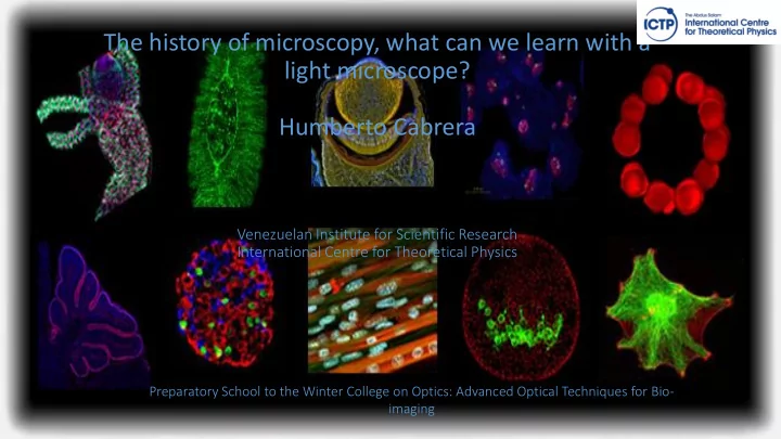
SLIDE 1 The history of microscopy, what can we learn with a light microscope? Humberto Cabrera
Venezuelan Institute for Scientific Research International Centre for Theoretical Physics
Preparatory School to the Winter College on Optics: Advanced Optical Techniques for Bio- imaging
SLIDE 2
Microscopy is often what first captivates kids with science
SLIDE 3
What the Telescope has done for studies of the universe
SLIDE 4
The microscope has done for biology
S2 cell anaphase
SLIDE 5
Microscopes allow us to explore beautiful worlds
Stephen J Smith - http://www.ncbi.nlm.nih.gov/pmc/articles/PMC 2693015/
SLIDE 6
SLIDE 7
“You can observe a lot just by watching” Yogy Berra
SLIDE 8
Microscopes reveal the dynamics of biological systems
Immune cells in a lymph node Philipe Bousso
SLIDE 9 Microtubules and F-actin, newt lung epithelial cell
Microscopes reveal the dynamics of biological systems
Drosophila embryo mitosis
SLIDE 10
Robert Hooke´s cell from cork 1665
SLIDE 11
Anton van Leeuwenhoek´s “Animalcules”, 1676
SLIDE 12
Walther Flemming pioneer of mitosis, 1878
SLIDE 13
Camillo Golgi´s silver staining of internal membranes (Golgi apparatus), 1898
SLIDE 14
Ramon y Cajals´cerebellar neurons, 1905
SLIDE 15
Shinya Inoue turns to live cell imaging
Mitosis in pollen mother cells from easter lilly 1951
SLIDE 16
Figure from H. Huxley and J. Hanson, Nature 1954 Hugh Huxley´s and Andrew Huxley´s studies of muscle contraction
SLIDE 17 How are proteins and membranes transported in nerve cells?
In 1960-70s, axonal trasport was studied primarily by following the movement of radioactively labelled proteins
SLIDE 18
A revolution in microscopy at the Marine Biological Laboratory: the birth of video microscopy
SLIDE 19
Video-DIC microscopy of squid giant axon, Allen, Brady Lasek, 1982
SLIDE 20 Purified kinesin moving artificial beads along microtubules, 1984 (Ron Vale) https://valelab.ucsf.edu/
Watching biochemistry in action
SLIDE 21 Shalfie, Shimomura and Tsien Nobel prize in 2008
Fluorescent Proteins Start a New Revival in Microscopy
SLIDE 22
Mic icroscopy is is constantly advancin ing
SLIDE 23
Resolution Lim imits of Lig ight
SLIDE 24 Breaking Resolution Barriers Super-resolution Microscopy
Xu K, Babcock HP, Zhuang X, Nature Methods 2012
SLIDE 25
Breaking Resolution Barriers Super-resolution Microscopy
Comparison of the resolution obtained by confocal laser scanning microscopy (top) and 3D structured illumination microscopy (3D-SIM- Microscopy, bottom). Shown are details of a nuclear envelope. Nuclear pores (anti-NPC) red, nuclear envelope (anti-Lamin) green, chromatin (DAPI-staining) blue. Scale bar: 1µm
SLIDE 26 Manip ipula lations of obje jects, mole lecules and cells lls wit ith lig light
Dance of beads Stretching RBCs by optical tweezers. (a) Two diametrically opposed silica beads
- f 4.1 μm are attached onto an RBC
- surface. (b) One bead is trapped by
- ptical tweezers while the other is fixed
- nto a glass surface. Deformation is
achieved by moving the glass surface to the opposite direction. (c) Large deformations of RBCs in phosphate buffer saline solution at room temperature are captured by optical micrographs under different trapping forces
- H. Zhang and K Liu, J. R. Soc. Interface (2008) 5, 671–690
SLIDE 27
Microscopy is making breakthroughs at all scale of biology
SLIDE 28
Measurements of sin ingle le mole lecule les
SLIDE 29
Measurements of sin ingle le mole lecule les
SLIDE 30
We acknowledge Profesor Ron Vale for the material used during the preparation of the lecture https://valelab.ucsf.edu/ https://www.ibiology.org/ibioeducation/taking-courses/ibiology-microscopy-course.html
SLIDE 31
Thanks