
SLIDE 1
ÓRgÃO OFICIAL DA SOCIEDADE PORTUgUESA DE REUMATOLOgIA 155
IMAgES IN RHEUMATOLOgy
Active and passive shoulder range of motion showed limited abduction (30º and 60º, respectively) and Jobe’s test was positive. Blood tests were unremarkable, show- ing a C-reactive protein of 0.56 mg/dL and erythrocyte sedimentation rate of 17 mm. Plain radiography re- vealed soft tissue swelling above the ACJ and elevation
- f the humeral head with GH joint space narrowing
and reduction of the subacromial space (Figure 2). Ul- trasound showed a loculated hypo-anechoic mass over- lying the ACJ with degenerative changes of the joint (Figures 3A and 3B), complete supraspinatus tear (Fi
- gure 3C), and GH synovitis and osteoarthritis (Figure
3D); no SASD bursitis was depicted. Aspiration of the ACJ was performed revealing 5 cc of yellow translucent gelatinous fluid with sparse hematic content; on po- larized light microscopy no crystals were found and bacterial cultures were negative. The diagnosis of supraspinatus tear, complicated by the formation of an ACJ cyst with “Geyser sign”, was made. The patient was
- 1. Serviço de Reumatologia e Doenças Ósseas Metabólicas,
Hospital de Santa Maria, CHULN, Centro Académico de Medicina de Lisboa, Portugal
- 2. Unidade de Investigação em Reumatologia, Instituto de
Medicina Molecular, Faculdade de Medicina, Universidade de Lisboa, Centro Académico de Medicina de Lisboa, Portugal
The geyser sign: an unusual presentation of a rotator cuff tear
Martins P1,2, Teixeira V1,2, Saraiva F1, Ponte C1,2
ACTA REUMATOL PORT. 2019;44:155-157
IntRodUctIon Acromioclavicular joint (ACJ) cysts are an uncommon clinical entity that may occur as consequence of full- thickness rotator cuff tears and degenerative ACJ
- changes. The “Geyser sign” is an unusual imaging sign
that derives its name from its geyser-like appearance.1,2 It occurs when synovial fluid from the glenohumeral (GH) joint extravasates through a full-thickness tear of the rotator cuff into the subacromial subdeltoid (SASD)
- bursa. Subsequently, the synovial fluid “erupts” through
a degenerated ACJ causing distension of its superior capsule and formation of a cystic mass. Two types of ACJ cysts have been described: type 1 limited to the ACJ and characterized by no communication with the GH joint, and type 2 defined by a rotator cuff tear which allows for a fluid communication to be established be- tween the ACJ and GH joint.3 Treatment can be man- aged nonoperatively or operatively, depending on the patient’s symptoms, age, medical comorbidities, and the presence of rotator cuff tear.3-5 casE PREsEntatIon A 76-year-old male presented to the emergency de- partment with a 1-week history of pain and swelling
- ver the superior aspect of the right shoulder. In addi-
tion, in the year preceding presentation, he had been complaining of limitation of motion and gradual pain
- f the GH joint. He denied constitutional symptoms,
such as fever or weight loss, or history of recent trau-
- ma. On inspection, he was found to have an important
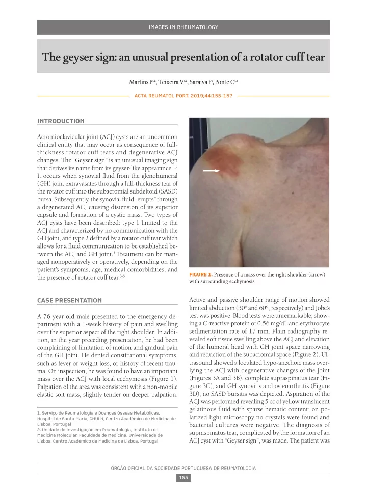
mass over the ACJ with local ecchymosis (Figure 1). Palpation of the area was consistent with a non-mobile elastic soft mass, slightly tender on deeper palpation.
FIGURE 1. Presence of a mass over the right shoulder (arrow) with surrounding ecchymosis