
SLIDE 1
3/26/2013 1
Surgical Problems in Primary Care
Ronald H. Labuguen, MD
Associate Clinical Professor UCSF Department of Family and Community Medicine
- UCSF Family Medicine Board Review Course
March 26, 2013
Surgical Problems in Primary Care: Stories from My Life
Ronald H. Labuguen, MD
Associate Clinical Professor UCSF Department of Family and Community Medicine
- UCSF Family Medicine Board Review Course
March 26, 2013
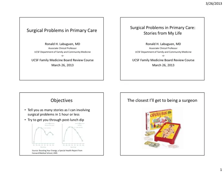
Objectives
- Tell you as many stories as I can involving
surgical problems in 1 hour or less
- Try to get you through post-lunch dip
Source: Boosting Your Energy, a Special Health Report from Harvard Medical School, 2005