surgical and non surgical
play

Surgical and Non-Surgical Adenomas Approaches for Large Pituitary - PDF document

Overview Large Pituitary Surgical and Non-Surgical Adenomas Approaches for Large Pituitary 1. Definitions, Epidemiology, and Clinical Masses Presentations 2. Surgical Management Manish K. Aghi , M.D., Ph.D. Professor 3. Postoperative


  1. Overview – Large Pituitary Surgical and Non-Surgical Adenomas Approaches for Large Pituitary 1. Definitions, Epidemiology, and Clinical Masses Presentations 2. Surgical Management Manish K. Aghi , M.D., Ph.D. Professor 3. Postoperative Results and Adjuvant Radiation Director, Center for Minimally Invasive Skull Base Surgery 4. Etiology California Center for Pituitary Disorders 5. Investigational Molecular Therapies Department of Neurosurgery University of California, San Francisco (UCSF)
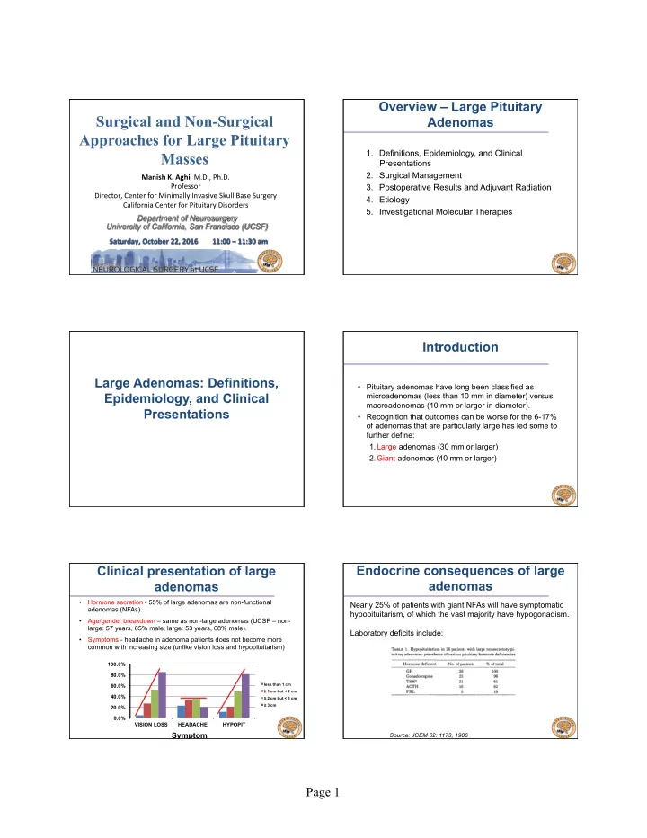
 Saturday, October 22, 2016 11:00 – 11:30 am Introduction Large Adenomas: Definitions, • Pituitary adenomas have long been classified as Epidemiology, and Clinical microadenomas (less than 10 mm in diameter) versus macroadenomas (10 mm or larger in diameter). Presentations • Recognition that outcomes can be worse for the 6-17% of adenomas that are particularly large has led some to further define: 1. Large adenomas (30 mm or larger) 2. Giant adenomas (40 mm or larger) Clinical presentation of large Endocrine consequences of large adenomas adenomas • Hormone secretion - 55% of large adenomas are non-functional Nearly 25% of patients with giant NFAs will have symptomatic adenomas (NFAs). hypopituitarism, of which the vast majority have hypogonadism. • Age/gender breakdown – same as non-large adenomas (UCSF – non- large: 57 years, 65% male; large: 53 years, 68% male). Laboratory deficits include: • Symptoms - headache in adenoma patients does not become more common with increasing size (unlike vision loss and hypopituitarism) 100.0% 80.0% less than 1 cm 60.0% ≥ 1 cm but < 2 cm 40.0% ≥ 2 cm but < 3 cm ≥ 3 cm 20.0% 0.0% VISION LOSS HEADACHE HYPOPIT Symptom Source: JCEM 62: 1173, 1986 Page 1

  2. Microscopy vs. Endoscopy – UCSF results re: NFA size inflection point • No difference in GTR rates was noted between the approaches Surgical Management of • Correlating GTR rates with size revealed the SI dimension to be where an inflection point occurred. Large Adenomas • This inflection point was smaller with the microscopic approach (3.1 cm – GTR rate if SI<3.1 cm=65%, GTR rate if SI ≥ 3.1 cm=32%) than with endoscopic cases (4.0 cm – GTR rate if SI<4.0 cm =59%, GTR rate if SI ≥ 4.0 cm=28%) Transcranial Approaches for Large Crani for pituitary tumors – anatomic Adenomas considerations influencing approach Transcranial approaches needed for giant adenomas that violate 10% chiasm over 80% chiasm over 10% chiasm over arachnoid planes and leave tumor in locations difficult to access tuberculum=prefixed diaphragm dorsum=postfixed endonasally (sylvian fissure, corpus callosum, third ventricle) IIIrd Vent 1. Interhemispheric 2. Subfrontal (uni- or 3. Pterional +/-orbital Dorsum bilateral) bar removal sellae Ant clinoid Two implications of the anatomy on pituitary adenomas – (1) affects the type of visual field defect (contralateral HH, bitemporal HH, and monocular deficit); (2) influences choice of craniotomy (avoid subfrontal for prefixed chiasm) Rate of GTR and approach selection for macro/large/giant adenomas Postoperative results UCSF experience – 721 adenomas 2007-2012 and adjuvant therapy 1. Macroadenomas (10-29 mm diameter) – 411 cases (57%), all for large resected endonasally, 85% GTR 2. Large adenomas (30 mm or larger) – 79 cases (11%), all nonfunctional resected endonasally, 75% GTR adenomas 3. Giant adenomas (40 mm or larger) – 50 cases (7%): 40 endonasal resections, 8 staged endonasal/craniotomy approaches, and 2 pure craniotomies. Of craniotomies: 5 orbitozygomatic, 3 subfrontal, 2 interhemispheric. 44% GTR Page 2

  3. Case – staged transsphenoidal and Case – staged transsphenoidal and transcranial approaches transcranial approaches 33 year old male with 3 years of right eye blindness that he was 62 year old female with over 15 years of right eye blindness and told was “ optic atrophy ” presents with headache temporal field cuts in left eye, presents with headache. Labs reveal hypopituitarism. Inter- Orbito- Trans- hemispheric Trans- zygomatic sphenoidal Transcranial sphenoidal Transcranial Giant adenoma seen through a Radiation for large pituitary transsylvian approach adenomas While no randomized trial has investigated postoperative radiation for giant NFAs, several retrospective series have shown benefit. The size of the targets in subtotally Temporal Frontal resected large or giant adenomas sometimes requires radiation therapy rather than radiosurgery. RadiaAon therapy Non-irradiated Source: Neurosurgery 60: 993, 2007 Postoperative endocrine Giant adenoma recurrence – UCSF improvement in large NFA patients experience • 31 giant adenomas compared to 66 non-giant Reported rates of recovery - 15% (GH), 30% (testosterone), 40% adenomas. (adrenal), and 50-60% (thyroid). Serum TSH • Rate of complete resection – 80% (non-giant) vs. 55% insulin-induced responses to (giant) (p=NS). hypoglycemia TRH • No completely resected adenomas recurred during short Source: postop follow-up, while progression rates for subtotally resected postop JCEM 62: Mean adenomas did not differ in giant vs non-giant. 1173, 1986 corAsol preop TSH postop Mean preop glucose preop Page 3

  4. Endocrine recovery after surgery for large NFAs (UCSF series) • UCSF experience – Of 129 giant/large adenomas over 5 years, 125 had documented hypopituitarism Etiology of Large preoperatively. • Rates of correction 6 weeks after surgery without Adenomas hormone replacement were 20% for testosterone, 36% for thyroid, and 29% for cortisol. • No patients with preoperative TSH below 0.03 (units), testosterone below 2.0 (units), or cortisol below 1 µ g/dL was able to be corrected sufficiently with surgery to avoid hormone replacement. Three possible etiologies of giant i. Different biology? - MIB-1 labeling adenomas in large vs. non-large adenomas i. start out with The UCSF experience 2005-2008 different biology 3.0% than smaller Giant adenomas 2.0% MIB-1 labeling ii. result from longer 1.0% Normal Gland Non-giant Giant incubation periods 0.0% < 30 mm 30 mm or larger iii. result from a second hit in a Giant smaller adenoma i. Different biology? - Atypical i. Different biology? – p53 Adenomas polymorphism • Performed full sequencing of the p53 gene in 35 pituitary adenomas • In 2004, WHO revised classification of pituitary adenomas • Polymorphism rs1042522:C>G in codon 72 of exon 4 whose C included an “ atypical ” variant with variant produces a proline and occurs in 64% of the population, has 1. MIB-1>3% a G variant producing an arginine in 62% of adenomas 2. excessive p53 immunoreactivity (p=1.09x10 -9 ). 3. increased mitoses. • The G variant renders adenomas more proliferative and causes • In our UCSF series, atypical adenomas were more invasive but patients to present a decade earlier with symptoms. not larger. We also found atypical adenomas to recur more frequently, but conversion from non-atypical to atypical did not 0.002 0.0018 G allele carriers occur. 0.0016 Probability General populaAon 0.0014 0.0012 0.001 0.0008 0.0006 0.0004 0.0002 0 Under 35 to 40 to 45 to 50 to 55 to 60 to 65 to 70 to 75+ 34 39 44 49 54 59 64 69 74 years Age Page 4

  5. ii. Longer incubation period? - ii. Socioeconomic factors leading to Factors leading to delayed diagnosis delayed diagnosis Unfortunately elderly patients with prolonged duration of visual Given lack of proliferative differences could longer adenomas symptoms are unlikely to return to baseline vision after surgery, just arise from prolonged incubation (i.e. delay in particularly when elderly diagnosis?) In the UCSF series, the greatest delay in being diagnosed 60% with adenoma in patients with visual symptoms occurred in Percent of patients elderly non-white patients who had a delay from onset of with 40% visual symptoms to adenoma diagnosis of over 6 months postop compared to 2 months in younger white patients). return to 20% baseline Duration of Delayed diagnosis in patients with visual symptoms often due vision n=32 over 6 months 0% visual to not seeking care or being diagnosed with other 6 or fewer months n=33 symptoms Age20s-30s conditions (cataracts, retinopathy, glaucoma), leading to a Age 40s-50s Age 60s-80s greater incidence of presenting with large adenomas. n=15 n=24 n=26 Age at diagnosis Source: Journal of Neurosurgery 116:283, 2012 Source: Journal of Neurosurgery 116: 283, 2012 iii. A second hit? – Ongoing studies • Site-directed biopsies have uncovered the Investigational Agents Studied regional heterogeity of malignant tumors • Preliminary work has suggested similar genetic Pre-Clinically For Large heterogeneity in pituitary adenomas Adenomas Investigational drugs for large Investigational drugs for large pituitary adenomas pituitary adenomas 2. Targeting Rb inactivation 1. Protein kinase C inhibitors • Rb (retinoblastoma) pathway PKC activity increased in adenomas; hypericin alterations seen in 75-90% of adenomas. Aberrantly activated (PKC inhibitor) inhibits proliferation of adenoma Rb/E2F1 pathway releases E2F1 cell lines to induce PTTG1 (pituitary tumor transforming gene), resulting in chromosome instability and proliferation. • Doxorubicin targets Rb-deficient cells (preclinical) • CDK2 inhibitor R-roscovit for Rb- hyperphosphorylated adenomas Source: Journal of Neurosurgery 85: 329, 1996 (clinical trial ongoing) Source: Mol Endocrinol 23: 2000, 2009 Page 5

Download Presentation
Download Policy: The content available on the website is offered to you 'AS IS' for your personal information and use only. It cannot be commercialized, licensed, or distributed on other websites without prior consent from the author. To download a presentation, simply click this link. If you encounter any difficulties during the download process, it's possible that the publisher has removed the file from their server.

Recommend


More recommend