spontaneous rupture of the bile duct associated with
play

Spontaneous Rupture of the Bile Duct Associated with Pancreatitis. A - PDF document

JOP. J Pancreas (Online) 2011 Mar 9; 12(2):149-151. CASE REPORT Spontaneous Rupture of the Bile Duct Associated with Pancreatitis. A Rare Presentation Mahesh K Goenka, Bhaswati C Acharyya, Pradeepta K Sethy, Usha Goenka Institute of


  1. JOP. J Pancreas (Online) 2011 Mar 9; 12(2):149-151. CASE REPORT Spontaneous Rupture of the Bile Duct Associated with Pancreatitis. A Rare Presentation Mahesh K Goenka, Bhaswati C Acharyya, Pradeepta K Sethy, Usha Goenka Institute of Gastroenterology, Apollo Gleneagles Hospital. Kolkata, India ABSTRACT Context Spontaneous rupture of the bile duct, although rare, has been described as a known surgical cause of jaundice in infancy after biliary atresia. Case report This article describes a four-year-old girl who presented with severe abdominal pain and features suggestive of acute pancreatitis, who developed gradual distension of the abdomen, and was found to have a ruptured bile duct, producing biliary peritonitis. She was managed with laparoscopic drainage of the peritoneal cavity. However, in view of the persistent biliary drainage, an ERCP was performed followed by stent placement for a bile duct leak. She was subsequently diagnosed as having a choledochal cyst. Conclusion A high index of suspicion, appropriate investigation, such as MRCP, combined with early drainage can help in reaching an early diagnosis, and reduced morbidity and mortality in this rare disorder. INTRODUCTION system (Figure 1). She was managed therapeutically as a case of acute pancreatitis. However, she continued to Spontaneous rupture of the bile duct is a rare clinical have pain and developed fever and distension of the situation. It is, however, a potentially fatal abdomen from the 4 th day of her admission. She was complication. The disease is more common in children found to be very toxic. Her leucocyte count rose to than in adults. Several mechanisms have been proposed 24,900 µL -1 with 96% neutrophils. Amylase and lipase regarding its etiology. We describe a child who had an on day 4 increased additionally to 1,300 IU/L and unusual presentation and was successfully managed 2,957 IU/L, respectively. Abdominal ultrasonography with laparoscopic drainage followed by ERCP. showed massive ascites and an ascitic fluid CASE REPORT examination showed it to contain bile with bile salts and bilirubin. Laparoscopic drainage of the ascitic fluid A 4-year-old girl who was otherwise well, presented at was carried out with a thorough peritoneal lavage with the emergency ward of a different centre in our city warm normal saline and a peritoneal drain was having had acute abdominal pain associated with inserted. Abdominal distension decreased but, even on obstipation for 3 days. She was found to be very ill, 10 th postoperative day, she continued to have a large anicteric and dehydrated. She was rehydrated with amount of bilious ascitic fluid draining from the fluids. Investigations revealed that her liver function was normal; however, her serum amylase level was 906 IU/L (reference range: 25-125 IU/L), and her lipase level was 451 IU/L (reference range: 22-51 IU/L); her total leucocyte count was 12,700 µL -1 (reference range: 4,000-10,000 µL -1 ) with predominant neutrophilia. Her CRP was 102 mg/dL (reference 
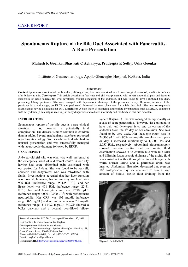
range: 0-6 mg/dL) and serum calcium was 7.5 mg/dL (reference range: 8.4-10.2 mg/dL). MRCP showed a bulky pancreas and a normal, non-dilated biliary Received November 11 th , 2010 - Accepted December 16 th , 2010 Key words Bile Ducts; Pancreatitis; Rupture Correspondence Mahesh Kumar Goenka Institute of Gastroenterology; Apollo Gleneagles Hospital; 58, Canal Circular Road; 700054 Kolkata; India Phone: +91-983-004-0599; Fax: +91-332-320-5218/2020 E-mail: mkgkolkata@gmail.com Document URL http://www.joplink.net/prev/201103/01.html Figure 1. Initial MRCP. JOP. Journal of the Pancreas - http://www.joplink.net - Vol. 12 No. 2 - March 2011. [ISSN 1590-8577] 149

  2. JOP. J Pancreas (Online) 2011 Mar 9; 12(2):149-151. Figure 2. Initial ERCP showing a leaking bile duct, biliary Figure 3. Subsequent ERCP showing a choledochal cyst. The stent sphincterotomy and stenting. has been removed. peritoneal drain. Fever persisted in spite of receiving whether acute pancreatitis precipitated the rupture or broad spectrum antibiotics. She was referred to our vice versa is difficult to say. A presentation of acute centre for further management. We performed an pancreatitis and the occurrence of bile duct perforation ERCP under anesthesia (Olympus TJF160 endoscope, after an attack of pancreatitis has rarely been described Singapore, Singapore) which showed evidence of a bile in children. Rupture of the right hepatic duct associated leak from the middle part of the common bile duct, with a choledochal cyst following acute pancreatitis, although the duct was not well delineated (Figure 2). however, has been described earlier in two children [4]. The intrahepatic radicles were, however, not dilated. Rupture of the bile duct is often associated with An endoscopic sphincterotomy was carried out and a pancreaticobiliary malunion with or without a biliary stent (7 Fr in diameter, 9 cm in length) was choledochal cyst [5, 6]. Both these disorders are known placed across the leak site into the left intrahepatic to have distal bile duct stenosis and intrinsic bile duct duct. wall weakness. In patients with pancreaticobiliary From the second post-ERCP day, her fever started to malunion, the junction of the pancreatic duct and the normalize The amount of bilious drain fluid decreased bile duct is situated outside the duodenal wall and, on day 5 after the ERCP, the peritoneal drainage containing the sphincter of Oddi [5]. Pancreatic juice tube was removed. Amylase and lipase levels became refluxing into the dilated bile duct is activated by normal and, after 7 days, the fever subsided. A repeat mixing with bile and causes acute inflammation and ultrasonography 2 weeks later was normal except for microabscess formation on the wall, eventually causing mild dilatation of the bile duct. ERCP was carried out 4 perforation. However, in a choledochal cyst, bile duct weeks later and the stent was removed. A rupture has been attributed to congenital mural cholangiogram showed a fusiform dilation of the upper weakness, usually at the junction of the cystic duct and two-thirds of the bile duct with no evidence of a leak the common bile duct, as well as to viral infections or (Figure 3). The intrahepatic biliary radicles were an ischemic etiology. The present case had a normal. A repeat MRCP also showed a choledochal choledochal cyst as the underlying mechanism. cyst (Figure 4). The child has been doing well and surgery for the choledochal cyst is planned for a later date. REVIEW OF THE LITERATURE Spontaneous bile duct perforation is not a common clinical entity. It was first described by Dijkstra in 1934 [1]. Since then, about 60 cases have been described in children [2]. It is most common in infancy, and occurrence as early as 25 weeks of gestation has been cited in the literature [3]. Median age at presentation is 4 years with a peak age of presentation at 6 months. The presenting symptoms are usually abdominal distension and/or jaundice. Our patient also had abdominal distension but was anicteric. Moreover, the present case had biochemical (raised amylase and lipase) and imaging evidence of acute pancreatitis; Figure 4. A repeat MRCP showing the choledochal cyst. JOP. Journal of the Pancreas - http://www.joplink.net - Vol. 12 No. 2 - March 2011. [ISSN 1590-8577] 150

Download Presentation
Download Policy: The content available on the website is offered to you 'AS IS' for your personal information and use only. It cannot be commercialized, licensed, or distributed on other websites without prior consent from the author. To download a presentation, simply click this link. If you encounter any difficulties during the download process, it's possible that the publisher has removed the file from their server.

Recommend


More recommend