an unusual presentation of a carcinoid tumor of the
play

An Unusual Presentation of a Carcinoid Tumor of the Common Bile Duct - PDF document

JOP. J Pancreas (Online) 2013 Jan 10; 14(1):85-87. CASE REPORT An Unusual Presentation of a Carcinoid Tumor of the Common Bile Duct Ashif Jethava 1 , Visvanathan Muralidharan 2 , Thalia Mesologites 3 , Elena Stoica-Mustafa 5 , Constantin A Dasanu


  1. JOP. J Pancreas (Online) 2013 Jan 10; 14(1):85-87. CASE REPORT An Unusual Presentation of a Carcinoid Tumor of the Common Bile Duct Ashif Jethava 1 , Visvanathan Muralidharan 2 , Thalia Mesologites 3 , Elena Stoica-Mustafa 5 , Constantin A Dasanu 4 Departments of 1 Hospital Medicine, 2 Gastroenterology, 3 Pathology, and 4 Hematology-Oncology, Saint Francis Hospital and Medical Center. Hartford, CT, USA. 5 Department of Pathology, Fundeni Clinical Institute. Bucharest, Romania ABSTRACT Context Carcinoid tumors arising from the bile ducts account for only a small fraction of biliary tract cancers. Case report We report herein a 42-year-old man with a carcinoid tumor of the common bile duct. He presented with abdominal pain, bloating and dyspepsia. Clinicolaboratory and imaging studies suggested a probable obstructive common bile duct lesion. The patient underwent an endoscopic retrograde cholangiopancreatography with a stent placement in view of common bile duct decompression. Persistence of symptoms prompted a laparotomy and pancreaticoduodenectomy that revealed a well-differentiated carcinoid tumor originating in the common bile duct. Conclusion Clinician’s familiarity with the unusual sites of origin of neuroendocrine tumors and/or atypical presentation of such tumors may facilitate their early recognition and allow for a timely intervention.
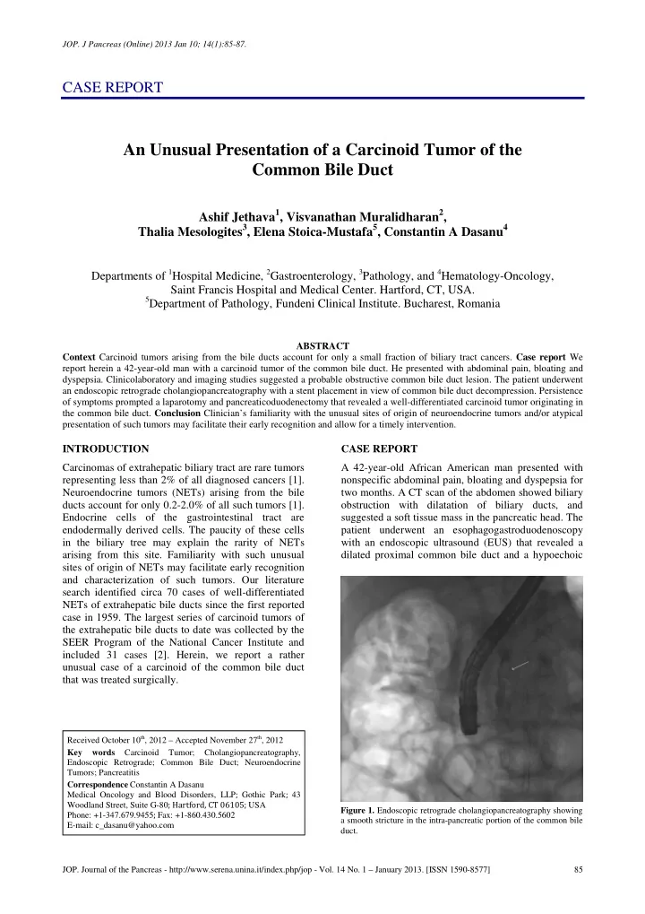
 INTRODUCTION CASE REPORT Carcinomas of extrahepatic biliary tract are rare tumors A 42-year-old African American man presented with representing less than 2% of all diagnosed cancers [1]. nonspecific abdominal pain, bloating and dyspepsia for Neuroendocrine tumors (NETs) arising from the bile two months. A CT scan of the abdomen showed biliary ducts account for only 0.2-2.0% of all such tumors [1]. obstruction with dilatation of biliary ducts, and Endocrine cells of the gastrointestinal tract are suggested a soft tissue mass in the pancreatic head. The endodermally derived cells. The paucity of these cells patient underwent an esophagogastroduodenoscopy in the biliary tree may explain the rarity of NETs with an endoscopic ultrasound (EUS) that revealed a arising from this site. Familiarity with such unusual dilated proximal common bile duct and a hypoechoic sites of origin of NETs may facilitate early recognition and characterization of such tumors. Our literature search identified circa 70 cases of well-differentiated NETs of extrahepatic bile ducts since the first reported case in 1959. The largest series of carcinoid tumors of the extrahepatic bile ducts to date was collected by the SEER Program of the National Cancer Institute and included 31 cases [2]. Herein, we report a rather unusual case of a carcinoid of the common bile duct that was treated surgically. Received October 10 th , 2012 – Accepted November 27 th , 2012 Key words Carcinoid Tumor; Cholangiopancreatography, Endoscopic Retrograde; Common Bile Duct; Neuroendocrine Tumors; Pancreatitis Correspondence Constantin A Dasanu Medical Oncology and Blood Disorders, LLP ; Gothic Park ; 43 Woodland Street, Suite G-80 ; Hartford, CT 06105; USA Figure 1. Endoscopic retrograde cholangiopancreatography showing Phone: +1-347.679.9455 ; Fax: +1-860.430.5602 a smooth stricture in the intra-pancreatic portion of the common bile E-mail: c_dasanu@yahoo.com duct. JOP. Journal of the Pancreas - http://www.serena.unina.it/index.php/jop - Vol. 14 No. 1 – January 2013. [ISSN 1590-8577] 85

  2. JOP. J Pancreas (Online) 2013 Jan 10; 14(1):85-87. Figure 2. A firm nodular mass measuring 1.7x1.3x0.8 cm in the common bile duct lumen. intraluminal density within the intrapancreatic portion of the common bile duct with post-acoustic shadowing. Subsequently, he underwent endoscopic retrograde cholangiopancreatography, which showed a smooth stricture in the intra-pancreatic portion of the common bile duct suggestive of an extrinsic compression (Figure 1). Cytology of the bile duct brushings was non-diagnostic, therefore, a biliary sphincterotomy with stent placement was performed. The patient presented a week later with worsening of upper abdominal pain. He had elevated transaminases, alkaline phosphatase, amylase and lipase times 2-4 the upper limit of normal, but normal serum bilirubin with fractions. A CT scan of abdomen and pelvis revealed an interval placement of a common bile duct stent with decompression of biliary system and a persistent prominence of pancreatic head. A magnetic resonance cholangiopancreatography showed a focal 80% narrowing of common bile duct with the presence of a stricture. The obstructive lesion was felt to be rather intraluminal or intramural than extrinsic in nature. The patient underwent a partial pancreaticoduodenectomy with gastrojejunostomy tube insertion. A white well- circumscribed, firm nodular mass measuring 1.7x1.3x0.8 cm was found at a distance of 3.3 cm from the common bile duct margin and 2.2 cm from the ampulla of Vater (Figure 2). The tumor was composed of cords or trabecullae of endocrine cells separated by Figure 3. Photomicrographs showing a well-differentiated common abundant fibrous stroma. The tumor cells had bile duct tumor positive for chromogranin and synaptophysin, monomorphic appearance and ample cytoplasm (Figure consistent with a carcinoid tumor. 3a). Surgical margins were clear and a total of nine peripancreatic lymph nodes were found to be negative biologic properties [3]. Ciaccio postulated the for metastases. Immunohistochemistry analysis revealed tumor cells positive for keratins, CD56, Ki- endocrine origin of these tumors in 1906 [3]. His findings were further reinforced by the work of Gosset 67, and negative for CK7. Staining for chromogranin and synaptophysin was positive, consistent with a and Masson in 1914 [3]. Davies was the first to carcinoid tumor (Figure 3bc). The postoperative course describe a carcinoid tumor of the common bile duct was uneventful and the patient was discharged from the and pancreatic duct in 1959 [4]. The most common anatomical locations for extrahepatic carcinoid tumors hospital. He remains disease-free six months postoperatively, with a good quality of life. are common bile duct (58%), perihilar duct (28%), cystic duct (11%) and common hepatic duct (3%) [5, DISCUSSION 6]. Our patient was diagnosed with a carcinoid arising First described by Lubarsch in 1888, carcinoid tumors from common bile duct that is the most common present characteristic histological, chemical and location for NETs of the extrahepatic biliary tract. JOP. Journal of the Pancreas - http://www.serena.unina.it/index.php/jop - Vol. 14 No. 1 – January 2013. [ISSN 1590-8577] 86

Download Presentation
Download Policy: The content available on the website is offered to you 'AS IS' for your personal information and use only. It cannot be commercialized, licensed, or distributed on other websites without prior consent from the author. To download a presentation, simply click this link. If you encounter any difficulties during the download process, it's possible that the publisher has removed the file from their server.

Recommend


More recommend