
SLIDE 1
Sarcoptes, Otodectes & Demodex Dr Lee Strapp BVetMed MRCVS - - PowerPoint PPT Presentation
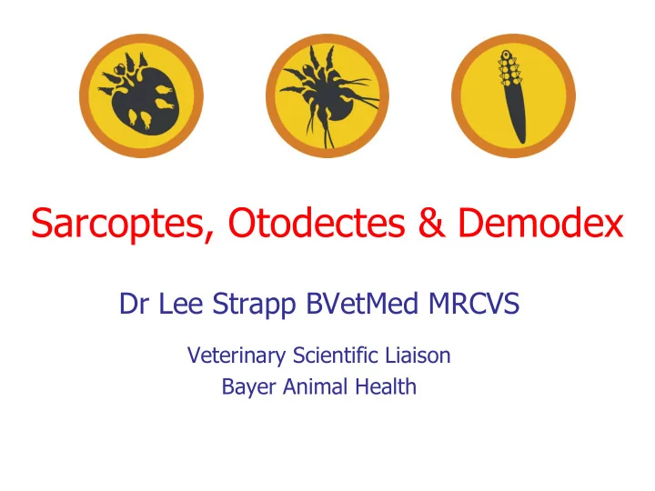
Sarcoptes, Otodectes & Demodex Dr Lee Strapp BVetMed MRCVS Veterinary Scientific Liaison Bayer Animal Health Overview Sarcoptes, Otodectes, Demodex Three different mites, all commonly encountered Obligate parasites - entire life
1 Egg 4 Tritonymph 2 Larva 5 Adult 3 Protonymph
Australian Veterinary Journal – Vol. 84, February 2006’ Fourie et al.
– physical presence of mites & mite saliva is an irritant
– Excoriation & wet eczema
day -1 day 112