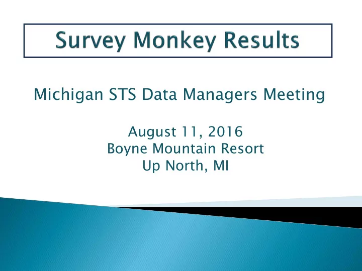
SLIDE 1
Michigan STS Data Managers Meeting
August 11, 2016 Boyne Mountain Resort Up North, MI
SLIDE 2 Present SM in a slightly different format to promote
“The Big Picture” scenario.
Ex
Expose e Ne Newer DM r DM’s to to Instructio ional Clin linical Scen cenarios…30 30% o
DM’s hav ave l les ess than an 1 1 year year of ex exper erien
e hav ave no no OR ex OR exposure.
Help to achieve statewide consensus by promoting
discussion and exchange.
SLIDE 3
- Reading the Scenario is necessary as the
format has changed from previous survey’s.
Thank You to those
that responded!!
- Please provide feedback regarding
this format in your evaluations!
- Rather than multiple scenarios, all questions
are based on a single scenario to mimic chart abstraction.
SLIDE 4
A 66 yo homeless male presents to the ER @ 8:00 pm. with unrelenting chest pain unlike any he has ever experienced before. He is hypotensive (BP in 70's), diaphoretic, ashen, agitated, and is demonstrating non- specific EKG changes. Unable to obtain an accurate H&P, and suspecting ACS, the patient is given ASA and Plavix, the STEMI team is notified, and he is taken directly to the Cath Lab. Imaging reveals normal coronary arteries, but identifies a 6 cm. ascending aorta, severe AI, "Severe" LV dysfunction, and pericardial tamponade. Further hemodynamic instability ensues, requiring intubation, and pericardiocentesis. Inotropes are started with no improvement in hemodynamics. The patient is immediately taken to the OR @ 9:15 pm. with a diagnosis of Aortic Dissection. Once on CPB, the surgeon discovers that the dissection originates in the ascending aorta, and extends past the aortic arch. The operation involves replacing the aortic valve with a 21mm bioprosthetic device sewn into a synthetic graft, attached distally at the Hemi-Arch, and re-implantation of the coronary ostia.
Patien ent S Scena enario
Part 1
SLIDE 5
Questi tion 1 n 1 Selec ections ns:
Prior MI = Yes (<=6 hrs.); Risk Factor's = Thoracic Dz; Pre-Op Meds = ASA + ADP Inhibitor + Inotropes; Cardiac Presentation at Surgery = STEMI. Prior MI = No; Risk Factor's = Thoracic Aorta Dz; Pre- Op Meds = ADP Inhibitor + Inotropes; Cardiac Presentation at Surgery = Other. Prior MI = Yes (<=6 hrs.); Risk Factor's = Unknown; Pre-Op Meds = ADP Inhibitor + Inotropes; Cardiac Presentation at Surgery = NSTEMI.
SLIDE 6 Poi
s to Con
er
Did the Patient have an MI?
What are his Risk Factor’s?
What Pre-Op Meds were administered? What is the Presentation at Surgery?
SLIDE 7 Poi
s to Con
er
Did the Patient have an MI?
What are his Risk Factor’s?
What Pre-Op Meds were administered? What is the Presentation at Surgery?
- No Cardiac Biomarker documentation
- “Non-Specific” EKG only
- No prior history available
SLIDE 8 Point nts t to Cons nsider er
Did the Patient have an MI? What are his Risk Factor’s? What Pre-Op Meds were administered? What is the Presentation at Surgery?
- Thoracic Dz. is only documented Risk Factor
- Documented: ASA, ADP Inhibitor, Inotropes
- NSTEMI, STEMI, Other
SLIDE 9 A patient presents for valve surgery with CHF, and no pain. Code other since the field is intended to capture cardiac presentation.
Tra raining M Manual E Exa xamples f for r “Other” r”
Patient does not need to be in active CHF to use “Other” as the cardiac presentation answer!
SLIDE 10 Survey s y says ays….. ..
Prio rior M MI = = Yes RF RF’s = = Th Thorac acic c Dz Dz Meds = = ASA+ADP+Inotr
Card rd Pre resent = = STEM EMI Prio rior M MI = = No RF RF’s = = Th Thorac acic c Dz Dz Meds = = ADP+Inotr
Card Pres esen ent = Other er Prio rior M MI =Y =Yes RF’s = = U Unknow
Meds = = ADP+Inotr
Car ard Present = NSTE NSTEMI
SLIDE 11 Questi tion
ections ns:
Previous Cardiac Intervention = Yes, Other Cardiac, Other; Heart Failure w/in 2 wks. = No; Cardiogenic Shock = Yes; Aortic Disease = Yes, Symptomatic, Hemodynamics Unstable. Previous Cardiac Intervention = Yes, Other Cardiac, Other; Heart Failure w/in 2 wks. = Yes, NYHA IV; Cardiogenic Shock = Yes; Aortic Disease = Yes, Location = Ascending. Previous Cardiac Intervention = No; Heart Failure w/in 2
- wks. = No, Cardiogenic Shock = Yes; Aortic Disease = Yes,
Lesion Type = Dissection.
SLIDE 12 Point nts t to Cons nsider er
Were there Previous Cardiac Interventions? Was there Heart Failure w/in 2 weeks? Was the Patient in Cardiogenic Shock? Aortic Disease: Presentation, Location, and Lesion Type?
- No documented Previous Interventions other than
Pericardiocentesis in Cath Lab.
SLIDE 13 Previous us C Cardiac I c Inter erven entions ns
- Pericardiocentesis is not a listed option for (#805) except for
Other Cardiac (not listed).
SLIDE 14
Cardi diac T Tampon ponade de
SLIDE 15
SLIDE 16 Point nts t to Cons nsider er
Were there Previous Cardiac Interventions? Was there Heart Failure w/in 2 weeks? Was the Patient in Cardiogenic Shock? Aortic Disease: Presentation, Location, and Lesion Type?
- Pericardiocentesis = Gray Area
- No consensus at the National level
- What is the Michigan consensus?
?
SLIDE 17
Heart F Failur ure e w/in 2 n 2 week eks
SLIDE 18 Point nts t to Cons nsider er
Were there Previous Cardiac Interventions? Was there Heart Failure w/in 2 weeks? Was the Patient in Cardiogenic Shock? Aortic Disease: Presentation, Location, and Lesion Type?
?
- Based on scenario,the patient does not fit the clinical
picture of HF
- No physician documented HF
SLIDE 19 Cardi diog
Shoc
- ck
- Sustained hypotension (BP 70’s)
- Inotropic support required.
SLIDE 20 Point nts t to Cons nsider er
Were there Previous Cardiac Interventions? Was there Heart Failure w/in 2 weeks? Was the Patient in Cardiogenic Shock? Aortic Disease: Presentation, Location, and Lesion Type?
?
- Meets the clinical definition of Cardiogenic
Shock
SLIDE 21 Ao Aort rtic D Dise isease
ntati tion
- n (most severe): Pain, hypotension, inotropes =
Symptomatic, Hemodynamically Unstable.
tion
- n (Choose all that apply): Where is the lesion present?
Ascending Aorta, Aortic Arch.
Type pe: : Dissection Tim imin ing: Acute Dis Dissectio ion T Type pe: : Stanford “Type A” ak aka: a: DeBakey “Type I”
SLIDE 22
SLIDE 23
Point nts t to Cons nsider er
Were there Previous Cardiac Interventions? Was there Heart Failure w/in 2 weeks? Was the Patient in Cardiogenic Shock? Aortic Disease: Presentation, Location, and Lesion Type?
?
SLIDE 24 Prev ev Card In Inter =Yes es, Other er Card Other er HF HF = = No Cardi dio
k =Yes Aortic ic Dz Dz = Yes, Sympt ptom
Prev ev Card In Inter =Yes es, Other er Card Other er HF HF = = Yes ess Cardi dio
k =No Aortic ic Dz Dz = Yes es, Ascen cending Prev ev Card rd Inter r =No HF HF = = No Cardi dio
k =Yes Aortic ic Dz Dz = Yes, Dissection
SLIDE 25 Questi tion
ections ns:
Aortic Valve Etiology = Primary Aortic Disease, Aortic Dissection; Aortic Valve Procedure Performed = Root Replacement w/ Valved Conduit; Aortic Procedure Location = Root, Ascending, and Hemi-Arch. Aortic Valve Etiology = Primary Aortic Disease, Aortic Dissection; Aortic Valve Procedure Performed = AVR and major root reconstruction with a valved conduit; Aortic Procedure Location = Root and Ascending. Aortic Valve Etiology = Primary Aortic Disease, Atherosclerotic Aneurysm; Aortic Valve Procedure Performed = AVR and insertion of non-valved conduit in a supra-coronary position; Aortic Procedure Location = Ascending and Hemi-Arch.
SLIDE 26
Point nts t to Cons nsider er
Aortic Valve Etiology Aortic Valve Procedure Aortic Procedure Location
SLIDE 27 Ao Aort rtic V Valve lve E Etio iology
- Aortic Insufficiency @ Cath found to be “Severe”
- Documented Aortic Dissection
SLIDE 28 Point nts t to Cons nsider er
Aortic Valve Etiology Aortic Valve Procedure Aortic Procedure Location
rimary ry A Aort rtic ic Dis Disease, , Aort rtic ic Dis Dissectio ion
SLIDE 29 Aortic V c Valve P Proced cedur ures es
(commercial or custom)
- Re-implant coronaries
- AVR
- Non-valved graft
in supra-coronary position
- AVR
- Root Reconstruction
- Valved conduit
Coro ronarie ies I Intact “Bentall”
SLIDE 30 Point nts t to Cons nsider er
Aortic Valve Etiology Aortic Valve Procedure Aortic Procedure Location
Primary ry Aort rtic D Dis isease, A Aort rtic D Dis issection
- The operation includes AVR, Root + Ascending Aorta
Replacement, Re-implantation of Coronary Ostia =
Ben Bentall
- What portions of the Aorta were involved in this procedure?
- Where did the Surgeon perform the distal anastomosis?
SLIDE 31
Aortic P c Proce cedur ure e Loca cation
SLIDE 32 Point nts t to Cons nsider er
Aortic Valve Etiology Aortic Valve Procedure Aortic Procedure Location
Primary ry Aort rtic D Dis isease, A Aort rtic D Dis issection
hat w was d done
- ne? The operation includes AVR, Root +
Ascending Aorta Replacement, Re-implantation of Coronary Ostia.
= Ben Bentall
- What portions of the Aorta were involved in this procedure?
- Where did the Surgeon perform the distal anastomosis?
Roo
t, A Asce cending A Aorta ta, H Hemi-Arc rch
SLIDE 33 Etiol
= Dissecti tion
Proced edure e = Benta tall Locatio ion = = Ascendin ing + + Hemi A i Arch Etiol
= Dissecti tion
Procedure = AVR+ R+ Ro Root Re Reco construct ct Location = n = Root + +Ascend nding ng Etiol
= Rupture Procedur ure = AVR+ S Supra c corona nary Cond ndui uit Locatio ion = = Ascendin ing + + Hemi A i Arch
SLIDE 34 After a prolonged surgery, the patient is transferred to ICU with considerable volume overload and marginal urine output. The surgeon
- rders UF for hemoconcentration. On the morning of POD #1, the patient
is found to be unresponsive, anuric, and has lost pulses in his lower
- extremities. MRI and CT Scans reveal a CVA, "ischemic" in origin, and a
new diagnosis of an abdominal aortic dissection. Urgent TEVAR is undertaken in the Vascular OR to repair the abdominal aorta with limited
- results. On POD #2, the patient remains unresponsive and anuric with
faint pulses in only one limb. UF has been replaced with CRRT due to rising creatinine. Repeat MRI reveals a worsened neurologic condition. On POD #3, the patient's condition is further complicated with worsening lactic acidosis and evidence of ischemic bowel. Given the poor prognosis, the family elects for palliative care measures only. The patient is extubated, and expires on POD #4.
Patien ent S Scena enario
Part t 2
SLIDE 35 Questi tion
ections ns:
Re-Op = Yes, Other Cardiac; CVA = Yes, Embolic; Renal failure = Yes, New Dialysis. Re-Op = Yes, Other Non-Cardiac; CVA = Yes, Undetermined Type; Renal failure = Yes, New Dialysis. Re-Op = Yes, Other Non-Cardiac; CVA = Yes, Hemorrhagic; Renal failure = Yes, New Dialysis. Re-Op = Yes, Other Cardiac; CVA = Yes, Undetermined Type; Renal failure = Yes, New Dialysis.
SLIDE 36 Point nts t to Cons nsider er
What at R Re-Opera ratio ion w was perform rmed p post-opera rativ ively ly? What t typ ype o
stroke d did the p pat atient su suffer? Did t the p patie ient d develo lop R Renal F l Failu ilure re?
- New diagnosis of abdominal dissection requires intervention.
- TEVAR is undertaken in Vascular Lab.
- Cardiac or Non-Cardiac (Vascular) ?
SLIDE 37
TEVA TEVAR
SLIDE 38 Point nts t to Cons nsider er
What at R Re-Opera ratio ion w was perform rmed p post-opera rativ ively ly? What t typ ype o
stroke d did the p pat atient su suffer? Did t the p patie ient d develo lop R Renal F l Failu ilure re?
- New diagnosis of abdominal (oops) dissection requires
intervention.
- TEVAR is undertaken in Vascular Lab.
- Cardiac or Non-Cardiac (Vascular) ?
- Embolic, Hemorrhagic, or Undetermined?
SLIDE 40
Po Post st-Op CVA CVA
SLIDE 41 Point nts t to Cons nsider er
What at R Re-Opera ratio ion w was perform rmed p post-opera rativ ively ly? What t typ ype o
stroke d did the p pat atient su suffer? Did t the p patie ient d develo lop R Renal F l Failu ilure re?
- New diagnosis of abdominal dissection requires intervention.
- TEVAR is undertaken in Vascular Lab.
- Cardiac or Non-Cardiac (Vascular) ?
- Embolic, Hemorrhagic, or Undetermined?
SLIDE 42
Renal F l Failu lure re
SLIDE 43
SLIDE 44
SLIDE 45 Point nts t to Cons nsider er
What at R Re-Opera ratio ion w was perform rmed p post-opera rativ ively ly? What t typ ype o
stroke d did the p pat atient su suffer? Did t the p patie ient d develo lop R Renal F l Failu ilure re?
- New diagnosis of abdominal dissection requires intervention.
- TEVAR is undertaken in Vascular Lab.
- Cardiac or Non-Cardiac (Vascular) ?
- Embolic, Hemorrhagic, or Undetermined?
- New requirement for postoperative dialysis
- Anuria >= 12 hours
SLIDE 46 Re ReOp=Yes es, O Other er C Card CVA = = Yes, E , Embolic ic RF RF= Y Yes Re ReOp=Yes es, O Other er NonCard CVA = A = Ye Yes, Undert rterm rmined RF RF= Y Yes Re ReOp=Yes es, O Other er NonCard CVA = = Yes, H Hemorrh rrhagic RF RF= Y Yes Re ReOp=Yes es, O Other er C Card CVA = = Yes, U Undet eter ermined ed RF RF= Y Yes
SLIDE 47 Questi tion
ections ns:
In-Hospital Post Operative Events include: Operative, Neurologic, Renal. In-Hospital Post Operative Events include: Pulmonary, Vascular, Other. All of the above.
SLIDE 48 Point nts t to Cons nsider er
- Intervention in Vascular Lab = Operative
- Post Operative Stroke (Undetermined) = Neurologic
- Ultra Filtration and new Dialysis = Renal
- Prolonged Ventilation = Pulmonary
- Acute Limb Ischemia = Vascular
- Aortic Dissection, GI Event, MSF(?) = Other
SLIDE 49 Questi tion
ections ns:
In-Hospital Post Operative Events include: Operative, Neurologic, Renal. In-Hospital Post Operative Events include: Pulmonary, Vascular, Other. All of the above.
SLIDE 50 Opera rativ ive Neuro rolo logic ic Re Renal al Pulmonary Vas ascu cular Other er All o
Above
SLIDE 51 Questi tion
ections ns:
The Mortality Cause of Death is: Cardiac The Mortality Cause of Death is: Neurologic The Mortality Cause of Death is: Vascular The Mortality Cause of Death is: Unknown The Mortality Cause of Death is: Deferred to Surgeon
SLIDE 52 Point nts t to Cons nsider er
- What was the Primary cause of death?
- What is the “most right” answer?
SLIDE 53
SLIDE 54
SLIDE 55 Questi tion
ections ns:
The Mortality Cause of Death is: Cardiac The Mortality Cause of Death is: Neurologic The Mortality Cause of Death is: Vascular The Mortality Cause of Death is: Unknown The Mortality Cause of Death is: Deferred to Surgeon
- Use POCMA as resource, discuss case, or call Jae
- Discuss case, ask questions, then call Jae
- Per TM, the Primary cause of all PO events
- Per POCMA, the seminal event that triggered the spiral
- Vascular events occurred as a result of initial insult
- Occurred as a result of initial surgery.
- Did not cause the cascade of events that followed
Seriously, please call us!!
SLIDE 56 Neuro Vascular Cardiac Unknown Surgeon