
SLIDE 1
925 Shergill Harbhajan et al., Int J Med Res Health Sci. 2015;4(4):925-927
Available online at: www.ijmrhs.com DOI: 10.5958/2319-5886.2015.00189.7 Case report Open Access
RARE PRESENTATION OF RUPTURED RUDIMENTARY HORN PREGNANCY
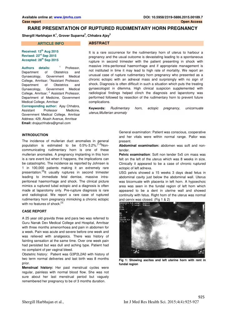
Shergill Harbhajan K1, Grover Suparna2, Chhabra Ajay3 INTRODUCTION The incidence of mullerian duct anomalies in general population is estimated to be 0.5%-3.2%.[1,2]Non- communicating rudimentary horn is one of these mullerian anomalies. A pregnancy implanting in this horn is a rare event but when it happens, the implications can be catastrophic. The incidence as reported by Johnsen is 1 in 100,000 patients making it an extremely rare presentation.[3]It usually ruptures in second trimester leading to immediate fetal demise, massive intra- peritoneal haemorrhage and shock. The clinical picture mimics a ruptured tubal ectopic and a diagnosis is often made at laparotomy only. Pre-rupture diagnosis is rare and radiological. We report a rare case of ruptured rudimentary horn pregnancy mimicking a chronic ectopic with no features of shock.[3] CASE REPORT A 25 year old gravida three and para two was referred to Guru Nanak Dev Medical College and Hospital, Amritsar with three months amenorrhoea and pain in abdomen for a week. Pain was acute and severe before one week and was relieved with analgesics. There was history of fainting sensation at the same time. Over one week pain had persisted but was dull and aching type. Patient had no complaint of per vaginal bleed. Obstetric history: Patient was G3P2L2A0 with history of two term normal deliveries and last birth was 8 months prior. Menstrual history: Her past menstrual cycles were regular, painless with normal blood flow. She was not sure about her last menstrual period but vaguely remembered her pregnancy to be of 3 months duration. General examination: Patient was conscious, cooperative and her vitals were within normal range. Pallor was present. Abdominal examination: abdomen was soft and non- tender. Pelvic examination: Soft non tender 5x5 cm mass was felt on the left of the uterus which was 8 weeks in size. Clinically it appeared to be a case of chronic ruptured ectopic of left adnexa. USG pelvis showed a 15 weeks 3 days dead fetus in abdominal cavity just below the abdominal wall. Uterus was bicornuate with placenta in left horn. A hypoechoic area was seen in the fundal region of left horn which appeared to be a dent in uterine wall and showed continuity with fetus. Right horn of the uterus was normal and cervix was closed. (Fig 1 & 2)
Fig 1: Showing ascites and left uterine horn with rent in fundal region
ABSTRACT
It is a rare occurrence for the rudimentary horn of uterus to harbour a pregnancy and the usual outcome is devastating leading to a spontaneous rupture in second trimester with the patient presenting in shock with massive intra-peritoneal haemorrhage and if appropriate management is not instituted in time it may lead to high rate of mortality. We report an unusual case of rupture rudimentary horn pregnancy who presented as a chronic ectopic with an adnexal mass and surprisingly with no sign of
- shock. Diagnosis is often difficult in such a situation which puts the treating
gynaecologist in dilemma. High clinical suspicion supplemented with radiological findings helped clinch the diagnosis and laparotomy was performed followed by resection of the rudimentary horn to prevent future complications. Keywords: Rudimentary horn, ectopic pregnancy, unicornuate uterus,Mullerian anomaly
ARTICLE INFO
Received: 13th Aug 2015 Revised: 23rd Sep 2015 Accepted: 28th Sep 2015 Authors details:
1
Professor, Department
- f
Obstetrics and Gynaecology, Government Medical College, Amritsar; 2Assistant Professor, Department
- f