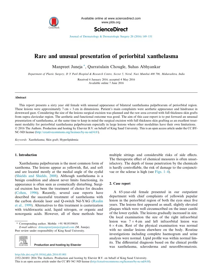

Available online at www.sciencedirect.com www.jdds.org ScienceDirect Journal of Dermatology & Dermatologic Surgery 20 (2016) 149–151 Rare and unusual presentation of periorbital xanthelasma Manpreet Juneja ⇑ , Qurratulain Chougle, Suhas Abhyankar Department of Plastic Surgery, D Y Patil Hospital & Research Centre, Sector 5, Nerul, Navi Mumbai 400 706, Maharashtra, India Received 6 January 2016; accepted 4 May 2016 Available online 7 June 2016 Abstract This report presents a sixty year old female with unusual appearance of bilateral xanthelasma palpebrarum of periorbital region. These lesions were approximately 7 cm � 3 cm in dimensions. Patient’s main complaints were aesthetic appearance and hindrance in downward gaze. Considering the size of the lesions surgical excision was planned and the raw area covered with full thickness skin grafts from supra clavicular region. The aesthetic and functional outcome was good. The aim of this case report is to put forward an unusual presentation of xanthelasma, at the same time to keep in mind the surgical excision with full thickness skin grafting as an excellent treat- ment modality for periorbital xanthelasma palpebrarum especially in large lesions where other modalities have their own limitations. � 2016 The Authors. Production and hosting by Elsevier B.V. on behalf of King Saud University. This is an open access article under the CC BY- NC-ND license (http://creativecommons.org/licenses/by-nc-nd/4.0/). Keywords: Xanthelasma; Skin graft; Hyperlipidemia 1. Introduction multiple sittings and considerable risks of side effects. The therapeutic effect of chemical measures is often unsat- Xanthelasma palpebrarum is the most common form of isfactory. The depth of tissue penetration by the chemicals xanthoma. The lesions appear as yellowish, flat, and soft is hardly controllable, the risk of damage to the conjuncti- and are located mostly at the medial angle of the eyelid vae or the sclerae is high (see Figs. 1–4). (Shields and Shields, 2008). Although xanthelasma is a benign condition and almost never limits functioning, its 2. Case report appearance is often seen as cosmetically disturbing. Surgi- cal excision has been the treatment of choice for decades A 65-year-old female presented in our outpatient (Cohen, 1996). Recently, several case reports have department with chief complaints of yellowish papular described the successful treatment of xanthelasma with lesion in the periorbital region of both the eyes since five the carbon dioxide laser and Q-switch Nd-YAG (Raulin years. The lesions first appeared as small, slightly elevated et al., 1999). Alternatives to this treatment is cauterization plaques which were well circumscribed on the inner canthi with trichloracetic acid, liquid nitrogen, or organic and of the lower eyelids. The lesions gradually increased in size. nonorganic acids. However, all of these methods bear On local examination the size of the right infraorbital lesion was 7 � 4 cm and left infraorbital lesion was ⇑ Corresponding author. Mobile: +91 9819359019. 6 � 4 cm. Rest of the physical examination was normal E-mail address: drmanpreetjuneja@gmail.com (M. Juneja). with no similar lesions elsewhere on the body. Routine Peer review under responsibility of King Saud University. investigations including complete haemogram and urine analysis were normal. Lipid profile was within normal lim- its. The differential diagnosis based on the clinical profile Production and hosting by Elsevier was xanthelasma, scleroderma and neurofibromatosis. http://dx.doi.org/10.1016/j.jdds.2016.05.001 2352-2410/ � 2016 The Authors. Production and hosting by Elsevier B.V. on behalf of King Saud University. This is an open access article under the CC BY-NC-ND license (http://creativecommons.org/licenses/by-nc-nd/4.0/).
150 M. Juneja et al. / Journal of Dermatology & Dermatologic Surgery 20 (2016) 149–151 Figure 1. Pre operative photograph of patient with bilateral periorbital Figure 4. Post operative photograph at 18 month follow up. xanthelasma in frontal view. vacuolated cytoplasm, a few lymphocytes and neutrophils in the dermis. Touton’s type giant cells were also noted. After thorough counselling of the patient and considering the cosmetic and functional complaints, we planned the surgical excision of the lesions and resurfacing of the defect with full thickness skin graft. The full thickness skin grafts were harvested from supraclavicular region to provide a good colour match. The check dressing was done after a period of 7 days. There was complete graft take. The patient was followed up to eighteen months with satisfac- tory outcome and no complications. 3. Discussion Xanthomas are lesions characterised by accumulation of Figure 2. Intraoperative photograph after excision of xanthelasma. lipid-laden macrophages. Xanthelasma palpebrarum is the most common of the xanthomas. A xanthelasma may instead be referred to as a xanthoma when it is larger and nodular, assuming tumourous proportions (Shields and Shields, 2008). It presents as an asymptomatic, usually bilaterally symmetric soft, velvety, yellow, flat, and polyg- onal papules around the eyelids. They are common in peo- ple of Asian origin and those from the Mediterranean region. Xanthelasmas are most common in the upper eyelid near the inner canthus. Xanthelasma may be associated with hyperlipidemia in which any type of primary hyper- lipoproteinemia can be present. Some secondary hyper- lipoproteinemias, such as cholestasis, may also be associated with xanthelasma. The colour of the patches and its appearance near the eyelids are enough to diagnose the condition. The diagnosis in a confounding case can be confirmed by tissue biopsy. Histologically, it is charac- terised by a regular epidermis and a dense distribution of Figure 3. Intraoperative photograph showing sutured full thickness graft. the xanthomitized macrophages interspersed by numerous Due to unusual appearance of the lesions, the definitive Touton’s type giant cells (Cohen, 1996; Breier et al., 2002). diagnosis was arrived at by tissue biopsy. The histopathol- Many people find this form of the condition particularly ogy showed localised collection of histiocytes with foamy embarrassing and disfiguring, hence opt for removal of the
Recommend
More recommend