
SLIDE 1
Radioimmunoassay of Biomolecule Based on the Lab-On-a-Chip Platform
Jin-Hee Kim a,b, So-Young Lee a and Seung-Kon Lee a*
aNeutron and Radioisotope Application Research Division, Korea Atomic Energy Research Institute,
111, Daedeok-daero 989beon-gil, Yuseong-gu, Daejeon 34057, Republic of Korea
bSchool of Architectural, Civil, Environmental and Energy Engineering, Kyungpook National University,
80, Daehak-ro, Buk-gu, Daegu 41566, Republic of Korea
*Corresponding author: seungkonlee@kaeri.re.kr
- 1. Introduction
RIA is an extremely specific and highly sensitive immunoassay, but the use of RIA is restrictive for the following reasons. RIA generally requires the long incubation time and this might result in the excessive radiation exposure of worker. The use of radioisotopes generates the radioactive wastes. Solid-phase RIA simplified the analysis procedures [1, 2], but it was not effective on the reduction of radioactive wastes and analysis time. The scintillation proximity assay (SPA) reduced the generation of radioactive wastes [3]. However, the reduction of analysis time remains an
- ngoing challenge.
Lab-On-a-Chip (LOC) platform is the miniaturized version of laboratory system. The LOC platform is composed of the microfluidic channel and it provides the advantages for bioassays such as short analysis time, less reagents consumption and automatic performance. The immunoassay based on the LOC platform reduces the reaction time, compared to the common
- immunoassay. It requires less reagents consumption, but
it sometimes has the problem of low signal intensity. We presented the RIA based on the microfluidic platform (μ-RIA) and optimized the reaction time for the analysis in previous study [4]. Here, we utilize the μ- RIA platform for the analysis of thyroxine (T4) antigen. The standard curve depending on the concentration of antigen is derived. The amount of antigen in QC sample
- f RIA kit is measured with the standard curve. Through
the experimental results, we show the applicability of μ- RIA for the analysis of biomolecules.
- 2. Experiments
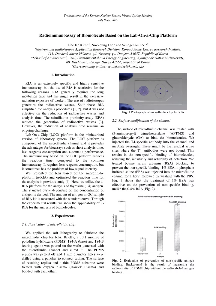
2.1. Fabrication of microfluidic chip We applied the soft lithography to fabricate the microfluidic chip for RIA. Briefly, a 10:1 mixture of polydimethylsiloxane (PDMS) 184-A (base) and 184-B (curing agent) was poured on the wafer patterned with the microfluidic channel and cured it. The PDMS replica was peeled off and 1 mm diameter holes were drilled using a puncher to connect tubing. The surface
- f resulting replica and a thin PDMS substrate were
treated with oxygen plasma (Harrick Plasma) and bonded with each other.
- Fig. 1 Photograph of microfluidic chip for RIA
2.2. Surface modification of the channel The surface of microfluidic channel was treated with (3-aminopropyl) trimethoxysilane (APTMS) and glutaraldehyde (GA) to bind the biomolecules. We injected the T4-specific antibody into the channel and incubate overnight. There might be the residual active sites where the T4 antibodies were not bound. This results in the non-specific binding of biomolecules, reducing the sensitivity and reliability of detection. We treated bovine serum albumin (BSA) blocking to prevent the non-specific binding. 1% BSA in phosphate buffered saline (PBS) was injected into the microfluidic channel for 1 hour, followed by washing with the PBS.
- Fig. 1 shows that the treatment of 1% BSA was
effective on the prevention of non-specific binding, unlike the 0.4% BSA (Fig. 2).
- Fig. 2 Evaluation of prevention of non-specific antigen
- binding. Background is the result of measuring the