
SLIDE 1
4/18/2015 1
PUT YOUR BEST FOOT FORWARD
Bala Ramanan, MBBS 1st year vascular surgery fellow
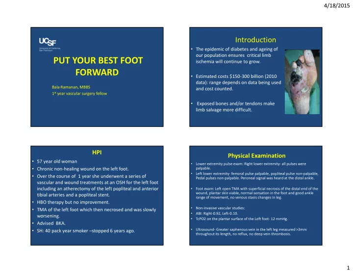
Introduction
- The epidemic of diabetes and ageing of
- ur population ensures critical limb
ischemia will continue to grow.
- Estimated costs $150-300 billion (2010
data): range depends on data being used and cost counted.
- Exposed bones and/or tendons make
limb salvage more difficult.
HPI
- 57 year old woman
- Chronic non-healing wound on the left foot.
- Over the course of 1 year she underwent a series of
vascular and wound treatments at an OSH for the left foot including an atherectomy of the left popliteal and anterior tibial arteries and a popliteal stent.
- HBO therapy but no improvement.
- TMA of the left foot which then necrosed and was slowly
worsening.
- Advised BKA.
- SH: 40 pack year smoker –stopped 6 years ago.
Physical Examination
- Lower extremity pulse exam: Right lower extremity- all pulses were
palpable.
- Left lower extremity- femoral pulse palpable, popliteal pulse non-palpable,
Pedal pulses non-palpable. Peroneal signal was heard at the distal ankle.
- Foot exam: Left open TMA with superficial necrosis of the distal end of the
wound, plantar skin viable, normal sensation in the foot and good ankle range of movement, no venous stasis changes in leg.
- Non-invasive vascular studies:
- ABI: Right-0.92, Left-0.10.
- TcPO2 on the plantar surface of the Left foot- 12 mmHg.
- Ultrasound- Greater saphenous vein in the left leg measured >3mm
throughout its length, no reflux, no deep vein thrombosis.