
SLIDE 1
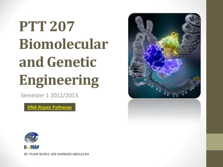
PTT 207 Biomolecular and Genetic Engineering
Semester 1 2012/2013
BY: PUAN NURUL AIN HARMIZA ABDULLAH
PTT 207 Biomolecular and Genetic Engineering Semester 1 2012/2013 - - PowerPoint PPT Presentation
PTT 207 Biomolecular and Genetic Engineering Semester 1 2012/2013 BY: PUAN NURUL AIN HARMIZA ABDULLAH We totally missed the possible role of enzymes in DNA repair. I later came to realize that DNA is so precious that probably many
Semester 1 2012/2013
BY: PUAN NURUL AIN HARMIZA ABDULLAH
We totally missed the possible role of enzymes in DNA repair…. I later came to realize that DNA is so precious that probably many distinct repair mechanisms would exist. Nowadays one could hardly discuss mutation without considering repair at the same time. Francis Crick, Nature (1974), 248:766
molecules.
by which a cell identifies and corrects damage to the DNA molecules that encoded its genome.
in its structure can result in mutations.
nucleotide sequence of DNA or from deletions, insertions, or rearrangements of DNA sequences in the genome.
induced.
Spontaneous mutations
e.g. DNA replication errors Induced mutations
an outside agent that causes DNA damage.
base with another, or one purine base with another.
with a purine or vice versa.
transversions is approximately 2:1
without changing the amino acid sequence are called synonymous mutations or silent mutations.
regions that do result in changed amino acids are called nonsynonymous mutations or missense mutations.
protein.
valine codon
stop codon is called a nonsense mutation.
protein synthesis.
not an exact multiple of three nucleotides, this results in a shift in the reading frame of the resulting mRNA.
nonfunctional protein.
Table : Codons (displayed as mRNA triplets)
EXPANSION OF TRINUCLEOTIDE REPEATS LEADS TO GENETIC INSTABILITY
conformations and unusual DNA secondary structures that interfere with transcription and DNA replication.
certain genetic neurological disorders.
Refer chapter 2.4 pg 31.
misaligns for recombination during meiosis with a different copy of the repeat in the homologous chromosome, instead of with the corresponding copy.
http://www.sci.sdsu.edu/~smaloy/MicrobialGenetics/topics/mutations/deletions.html
single- and double-strand breaks, 8- hydroxydeoxyguanosine residues, and polycyclic aromatic hydrocarbon adducts.
they can be correctly repaired if redundant information, such as the undamaged sequence in the complementary DNA strand or in a homologous chromosome, is available for copying.
be prevented, and, thus, translation into a protein will also be blocked. Replication may also be blocked and/or the cell may die.
sequence of the DNA.
is present in both DNA strands, and, thus, a mutation cannot be repaired.
function and regulation.
cells, mutant cells will increase or decrease in frequency according to the effects of the mutation on the ability of the cell to survive and reproduce.
mutations are related because DNA damages often cause errors of DNA synthesis during replication or repair; these errors are a major source of mutation.
causing a modification in one or more bases in a DNA sequence.
DNA bases.
an alkyl group (typically a small hydrocarbon side chain such as a methyl or ethyl group, denoted as-CH3 and-C2H5, respectively) to a DNA base.
atom binds to a carbon atom in the DNA base
cyclobutane ring between adjacent thymines, forming a T-T dimer.
block transcription and replication.
cytosine and thymine.
thymine-thymine dimer thymine-cytosine dimer
intercalating agents and base analogs:
rings that insert between the DNA bases.
mispair with guanine.
Formation of abasic sites
unstable base adducts. Double-stranded DNA breaks
chemical compounds.
the integrity and accessibility of essential information in the genome (but cells remain superficially functional when so-called "non- essential" genes are missing or damaged).
damage so that life can go on
structure without breaking backbone
Translesion synthesis (TLS)
polymerases transiently replace the replicative polymerases and copy past damaged DNA.
different compounds and types of radiation that can alter the chemical composition of the DNA. In response, the cell has developed different types of DNA repair mechanisms that can remove the lesion. When the lesion is not removed before replication is initiated, it can result in a block of the replication machinery that can ultimately lead to cell death. To bypass these blocks, specialized translesion synthesis (TLS) DNA polymerases are recruited to the site of the lesion. The TLS polymerases are capable of DNA synthesis over the damaged DNA, after which the replicative DNA polymerase can continue normal DNA synthesis. These TLS polymerases are generally error prone and have been implicated in drug resistance in bacteria and in different forms of cancer in humans.
Error-prone DNA polymerases
lesion: nucleotide substitution
DNA polymerase eta ()
Adenine residues.
dNTPs instead of one.
hold the TT dimer so that the two thymines can be paired with two adenines.
Reversal of thymine-thymine dimers by DNA photolyase
can be directly repaired.
light to break the covalent bonds holding two adjacent pyrimidines together.
DNA photolyase has two cofactors:
Damage reversal by DNA methyltransferase
in DNA induced by alkylating mutagens. This DNA adduct is removed by the repair protein, O6-methylguanine-DNA methyltransferase.
group on O6-methylguanine to the sulfhydryl group of a cysteine residue on the enzyme.
STRUCTURAL DISTORTIONS BY REMOVAL OF DNA DAMAGE
Base excision repair
due to conversion of one base to another.
the damaged base.
damage in a vast expanse of undamaged DNA?
STRUCTURAL DISTORTIONS BY REMOVAL OF DNA DAMAGE
alkylation.
called the "abasic site" or "AP site".
site and remove its base.
and neighboring nucleotides.
ligase.
STRUCTURAL DISTORTIONS BY REMOVAL OF DNA DAMAGE
Mismatch repair
result from DNA polymerase errors during replication.
excised.
mammalian cells is currently unknown.
STRUCTURAL DISTORTIONS BY REMOVAL OF DNA DAMAGE
Nucleotide excision
the damaged nucleotides (e.g., the dimer induced by UV light). The gap is then filled by DNA polymerase I and DNA
("RAD" stands for "radiation"), such as RAD3, RAD10. etc.
whole genome.
the transcribed strand of active genes.
reactive oxygen species, ionizing radiation, and chemicals the generate reactive oxygen species (free radicals).
nonhomologous end-joining.
Homologous recombination
information from an undamaged homologous chromosome. Nonhomologous end-joining (NHEJ)
ends without any requirement for sequence homology.
Thank You