SLIDE 1
10/24/2015 1
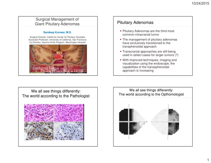
Sandeep Kunwar, M.D.
Surgical Director, California Center for Pituitary Disorders Associate Professor, University of California, San Francisco Co-Director, Gamma Knife Program, Washington Hospital
Surgical Management of Giant Pituitary Adenomas
Pituitary Adenomas
Pituitary Adenomas are the third most common intracranial tumor The management of pituitary adenomas have exclusively transitioned to the transphenoidal approach Transcranial approaches are still being used in select cases for larger tumors (?) With improved techniques, imaging and visualization using the endoscope, the capabilities of the transsphenoidal approach is increasing
We all see things differently: The world according to the Pathologist
We all see things differently: The world according to the Opthomologist
SLIDE 2
10/24/2015 2 We all see things differently: The world according to the Endocrinologist We all see things differently: The world according to the Endocrinologist/Neurosurgeon We all see things differently: The world according to the Neurosurgeon
Giant Pituitary Adenomas
What are Giant Pituitary Adenomas? Do giant adenomas arise from more aggressive adenomas or are they just a delay in diagnosis? Best management for these tumors?
SLIDE 3
10/24/2015 3
Giant Pituitary Adenomas
Definition:
¬ No general consensus to size ¬ Several studies (Cappabianca, et al,
Gondim, et al, Yang, et al, Goel, et al) defined this to be 4cm, while other large studies have defined this to be 3cm (Juraschka, et al)
Microadenoma (<1cm) Macroadenoma (>1cm) Large adenoma (>3 cm) Giant adenoma (>4 cm)
Giant Pituitary Adenomas
Retrospective analysis of the first consecutive 1000 endonasal transsphenoidal surgeries performed Surgeries performed 2001-2008 159 patients operated on had tumors >3 cm 59 patients had tumors >4cm
Giant Pituitary Adenomas
Ages ranged from 9-80 yo
¬ Mean age was 49 yo
Tumor sizes were 40-72mm
¬ Mean max tumor length was 45mm
41 M (69%), 18 F (31%) 7 patients had prior surgery
¬ 6 prior transsphenoidal surgery ¬ 1 prior transcranial surgery
Giant Adenomas
52 patients had Non-functioning adenomas (88%) 2 patients had acromegaly (3%) 2 patients had Cushing’s disease (3%) 3 patients had prolactinomas
¬ All 3 patients had failed medical therapy
(cabergoline)
SLIDE 4 10/24/2015 4
Giant Adenoma – Presenting Symptoms
Visual acuity loss was documented in 82% of patients Significant headaches were present in 17% of patients Diplopia was present in 5% of patients
Case Presentation 1 - Giant Adenoma (5.5 cm)
- 70 yo male with bitemporal vision loss, headache,
panhypopituitarism
Goals of therapy
Decompress optic nerves Decompress neural tissue (hypothalamus) Minimize neural trauma Minimize field of radiation therapy if needed
Case 1 - Outcome
Patient underwent extended endonasal approach with endoscopic assist Patient had marked improvement in vision He had transient postoperative DI, but at 6 wk follow-up was not on DDAVP Discharge from the hospital on POD#2 Pathology – pituitary adenoma with no atypical features
SLIDE 5
10/24/2015 5
Case 1 – Postop MRI scans (at 6 yr f/u pt had no recurrent disease)
Case Presentation - 2
51 yo F presented with vision loss Clinical appearance classic for acromegaly Hormonal work-up
¬ Prl – 55 ¬ GH – 10.9 ng/ml ¬ IGF-1 – 662 ng/ml
MRI showed a 4.7cm adenoma
Case 2 – MRI Goals of treatment
Decompress optic nerves Decrease/normalize IGF-1/GH Minimize neural trauma
SLIDE 6 10/24/2015 6
Case Presentation 2 - Outcome
Patient underwent extended endonasal transphenoidal surgery with GTR Patient was discharged on POD#1 At 12 week follow up
¬ GH – 1.1 ng/ml ¬ IGF-1 – 144 ng/ml ¬ Prolactin – 7
Pathology showed an atypical adenoma
¬ + for GH and Prolactin ¬ KI67 – 5% ¬ P53 – 5%
Case 2 - Follow-up MRI Case Presentation - 3
18 yo M with progressive vision loss and
¬ BTH and nasal field defect OD
At presentation he was noted to have DI and panhypopituitarism (prolactin nl) Pt with DM-2, morbid obesity, metabolic syndrome Patient also had hydrocephalus and a VP shunt was placed prior to referral
Case 3 - MRI scan
SLIDE 7
10/24/2015 7
Goals of treatment:
Decompress optic nerves Decompress hypothalamus Minimize radiation field
Case 3 - Treatment
Patient underwent extended endonasal transphenoidal surgery (2006) for subtotal resection of his tumor Postoperatively, his DI was difficult to manage and was discharged on POD#7 Vision improved OU Pathology showed atypical pituitary adenoma (KI-67 6%) Patient underwent radiation therapy 3 months after surgery
Case 3 - Follow-up MRI
MRI stable at 8 yr follow-up
Case Presentation - 4
58yo F with progressive vision loss
¬ Blind OD, ¾ defect OS with LP
No headaches Hormonal workup revealed normal prolactin with panhypopituitarism
SLIDE 8 10/24/2015 8
Case 4 - MRI scan
Tumor measured: 60x70x40 mm in size
Case 4 - Treatment
Goals:
¬ Decompress hypothalamus/Frontal
lobes
¬ Decompress Optic nerves ¬ Minimize neural trauma
Approach?
¬ Transcranial ¬ Transsphenoidal ¬ Both?
Case 4 -Treatment
Patient underwent extended endonasal transphenoidal surgery (2004) with resection of 80%
She was discharged to home on POD#2
¬ No DI
Her 3 month postop MRI showed a residual tumor in the cavernous sinus and suprasellar region, left optic nerve decompressed, right was decompressed but still distorted
¬ OS – finger counting ¬ OD – NLP
She underwent another endonasal transsphenoidal surgery at 6 months (2005) Pathology – pituitary adenoma, no atypia
Case 4 – Postop MRI
MRI 3/31/2014 – stable residual disease (no XRT)
SLIDE 9 10/24/2015 9
Giant Pituitary Adenomas - Complications
There were no deaths in this series Complications:
¬ Sinus infection: 14% ¬ CSF leak: 5% ¬ Permanent DI: 5% ¬ Carotid injury: 0% ¬ Stroke: 0%
Giant Pituitary Adenomas
Surgical Tips:
¬ Intraoperative navigation ¬ Use of lumbar subarachnoid drain to assist
in descent of suprasellar capsule
¬ Develop margins early and debulk centrally
to facilitate descent of suprasellar capsule
¬ Use of endoscope ¬ Use of a suction on suction technique to
tease capsule down
Giant Pituitary Adenomas - Conclusion
Treatment decision is based on goals
¬ Since majority of tumors present with vision
loss, surgery is warranted
¬ All patients must undergo hormonal and
- pthomalogical evaluation prior to treatment
including prolactin levels
¬ Prolactinomas should only be considered for
surgery if:
- They have failed medical therapy
- Have rapid onset of vision loss with
hemorrhage
- Develop a spontaneous CSF leak with
medical therapy
Giant Pituitary Adenomas - Conclusion
Transsphenoidal surgery is safe and effective in this population with low morbidity Allows rapid decompression of the optic nerves and hypothalamus Should only be considered if:
¬ tumor does not extend 1cm lateral to the ICA ¬ There are no vessels invaginating/wrapped into
the outer margins of the suprasellar tumor
Residual tumor may apoplex postop (particularly with “mickey mouse” ears)
SLIDE 10
10/24/2015 10
No transphenoidal surgery! Giant Pituitary Adenomas - Conclusion
In certain cases, complete resection may be possible
¬ No cavernous sinus invasion, smooth tumor
margins
Goals of surgery should be determined in advance
¬ Subtotal resection is ok ¬ Radiation therapy is an effective
postsurgical treatment
California Center for Pituitary Disorders
Department of Neurosurgery Manish Aghi Phil Theodosopolous Tarun Arora Gwen Stanhope Lewis Blevins Division of Endocrinology Lewis Blevins Blake Tyrell Division of Neuroradiology William Dillon Chris Hess Division of Neuropathology Andrew Bollen Tarik Tihan Airie Perry