
SLIDE 1
10/24/2015 1
Stereotactic Radiosurgery for Pituitary Adenomas
Jason Sheehan, MD, PhD Departments of Neurological Surgery And Radiation Oncology University of Virginia
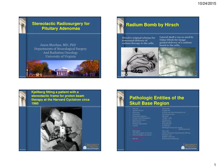
Radium Bomb by Hirsch
Hirsch’s original schema for transnasal delivery of radium therapy to the sella Lateral skull x-ray as used by Oskar Hirsh for image guided delivery of a radium bomb to the sella.
Kjellberg fitting a patient with a stereotactic frame for proton beam therapy at the Harvard Cyclotron circa 1960
Pathologic Entities of the Skull Base Region
- Abscess
Aneurysm
- Arachnoid Cyst
Cepahocele
- Chloroma
Colloid cyst/pars intermedia cyst
- Chordoma
Chondrosarcoma
- Craniopharyngioma
Dermoid
- Ectopic neurohypophysis
“Empty” sella
- Epidermoid tumor
Germinoma
- Hamartoma
Histiocytosis
- Pituitary hyperplasia
Hypophysitis
- Lipoma
Lymphoma
- Meningioma
Meningitis (bacterial, fungal, granulomatous)
- Metastasis
Mucocele
- Nasopharyngeal carcinoma
Opticochiasmatic-hypothalamic Glioma
- Osteocartilagenous tumors
Parasitic cyst
- Pituitary adenoma
Rathke’s cleft cyst
- Sarcoid
Esthesioneuroblastoma
- Schwannoma