
SLIDE 1
PICsl-FBI photonic- electronic Imaging Facility IBDM Imaging - - PowerPoint PPT Presentation
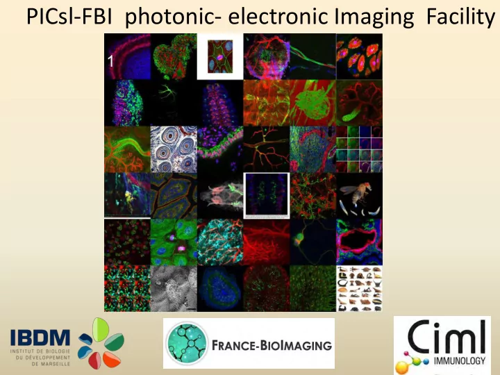
PICsl-FBI photonic- electronic Imaging Facility IBDM Imaging facility 200 researchers + 20 Quality process Service industrials Video-microscopy Tcn 11 video-microscopes scientific supervisor CNRS Pierre-Franois Lenne + DR2 CNRS 4
Technical supervisor
Cédric Matthews IR2 CNRS
Quality process Video-microscopy Tcn CNRS
Service 11 video-microscopes
Confocal Metrology
service 4 confocal, 3 spinning disk
machining/ microscopy AI CNRS
project computer-aided design / machining Two photon microscopy development Cédric Matthews Service / Project 2 two photon Image processing
scientific supervisor
Pierre-François Lenne DR2 CNRS
1 two photon , 1macro- confocal
200 researchers + 20 industrials + 4 Master Degree’s Training Operating Budget 2014 100 000 euros Capital Budget since 2009 1 million euros
1 IR , 1 AI , 1 TCN 22 systems
Confocal 2 LSM 510 meta 1 LSM 510 1 LSM 780 1 nikon macroconfocal Two photons 1 nikon high speed + script 1 zeiss 7 MP with 5 detectors 1 zeiss 510 NLO inverted Spinnind disk 1 Roper- yokogawa X1 with 2 cameras evolve 1 roper- yokogawa CSU 10 dedicated to electrophysiology Video microscopes 3 apotomes up to date 1 environnemental video-microscope 3 microscopes
Confocal LSM 510 UV meta upgrade Spinning disk (team Lecuit) Camera pixel size 8µm High resolution microscope STED Specific dyed antibodies Déconvolution Treatment Huygens software Omero database for images and assistance Technological transfert from Lenne team : PALM microscope
systems
dynamic imaging and contrast
processing and images analysis
deconvolution Prototype
(coming soon)
Associated to systems
publication
machining
IBDM training
Vocational training
control
videomicroscope confocal two photons STED
11 individual trainings on video-microscopes and 15 on other microscopes from January to June
spinning LSM 780 LSM 510 video STED
In the facility, we work as project
How we can help you ?
Which type of experiment, we choose
In collaboration with the team
EM and Team people
Help for image interpretation
In 2013 : 33 Projects in 2013 =>
57,58 42,42 External project Internal project
We are a facilities
Jean-Paul chauvin Aicha Aouane Fabrice Richard IE1 TCS IE2
We have projects with IBDM And many equipment TEM CIML SEM Timone Hospital Ultracryomicrotome Inmed AFS => cryossubstitution IMM HPF => cryofixation and much more in or out of PACA and more
Facility news :
question New microscope Existing equipment
Fusion of 2 platforms ImagImm is still a part of PICsL People
Didier Marguet & Philippe Pierre Supervisors Sophie Brustlein CDD (until Sept. 2014) Mathieu Fallet IE Sébastien Mailfert IR
Imaging Immunity
2 Two photon
+ temperature and anesthesia control Courtesy of M. Bajénoff’s lab
3 Widefield
+ temperature control Courtesy of M. Sieweke’s lab
3 Confocal
Courtesy of M. Dalod’s lab
3 Spinning disk
+ temperature control Courtesy of P. Golstein’s lab
4 Single molecule
+ temperature control Courtesy of H&M’s lab
from molecules to organisms
4 Single molecule
Bleaching Resolution Speed acq. Multicolor Depth (z) <30µm 1 low
3 Spinning disk
Bleaching Resolution Speed acq. Multicolor Depth (z) <30µm 2 excellent
3 Confocal
Bleaching Resolution Speed acq. Multicolor Depth (z) <30µm 4-8 high good good low medium Bleaching Resolution Speed acq. Multicolor Depth (z)
3 Widefield
<30µm 3 good medium Bleaching Resolution Speed acq. Multicolor Depth (z) <200 µm 2-3 medium
2 Two photon
medium medium good low excellent
from molecules to organisms
2013 2014 (January to May) Widefield microscopy 28% 20%↘ Confocal imaging (LSM780 + SP5) 78% (92% + 64%) 87%↗ (99% + 75%) Spinning disk (Perkin + Visitron + polar) 15% (Perkin) 22%↗ (33% + 14% + 20%)
Significant increase over the last years of the service by CIML’s teams
Technique 2013 2014 (January to June) Widefield microscopy NA 40 Confocal imaging NA 50 Spinning disk NA 10 Image Processing NA xxx
For fixed samples, multi-color imaging (up to 7 colors), high sensitivity
High content screening is a clear need to image large number of slides (service from the histology platform)
2013
in living cells – 13/19 mai 2013
19/22 novembre 2013
novembre 2013
20/24 octobre 2014
Intravital multiphoton microscopy (3 colors and CARS/SHG/THG)
Obtain more constrats in living animal imaging collaboration with Leica
Excitation-polarization-resolved confocal microscopy
Molecular order in living cell at video rate
collaboration with the MOSAIC group at Institut Fresnel
Super-resolution (STORM/PALM)
Molecular distribution and counting collaboration with the PhyTi group at Institut Fresnel
Multimodal FCS setup (FCCS/FLIM/STED)
Molecular interaction in living cells collaboration with ISS
20 40 60 80 100 120 140 160 180Y
CFP-CFP YFP-YFP Tom - Tom YFP - Tom CFP - Tom CFP - YFP