Page 1 Malignant Peripheral Nerve Sheath Tumor Protein Correlates - - PDF document

page 1
SMART_READER_LITE
LIVE PREVIEW

Page 1 Malignant Peripheral Nerve Sheath Tumor Protein Correlates - - PDF document

NEXT-GENERATION IMMUNOHISTOCHEMISTRY Disclosures FOR SOFT TISSUE TUMORS: FROM DIFFERENTIATION TO MOLECULAR GENETICS Dr. Jason Hornick discloses the following financial interests: Jason L Hornick, MD, PhD Director of Surgical Pathology


slide-1
SLIDE 1

Page 1

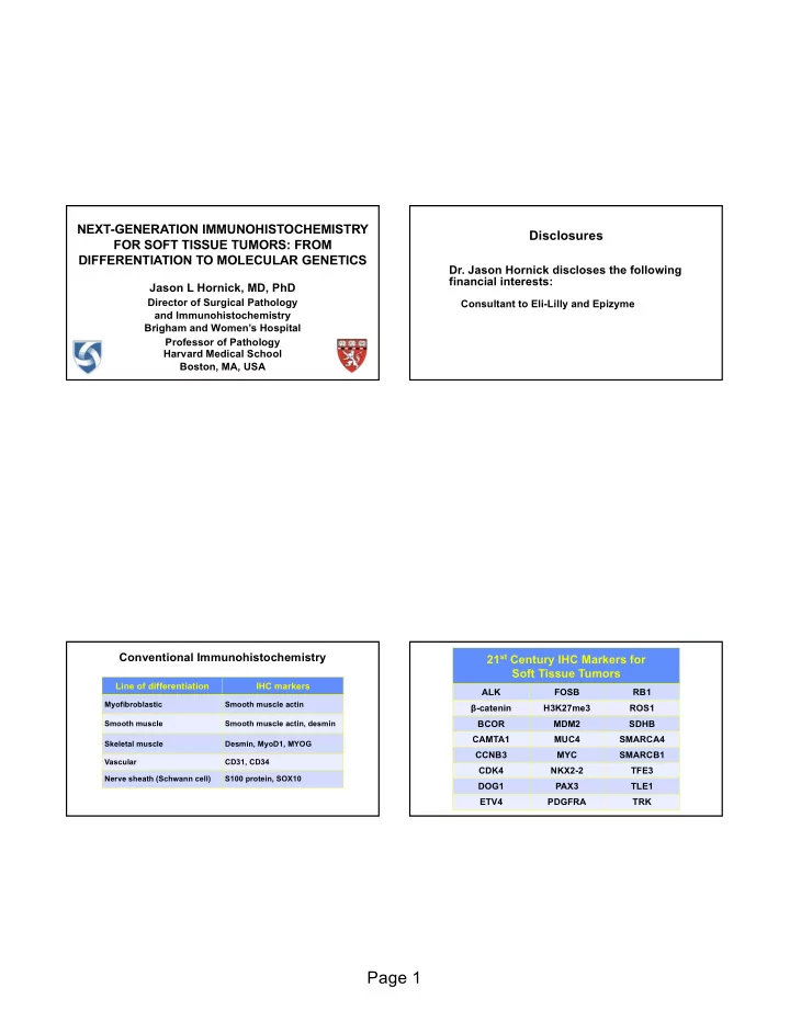
NEXT-GENERATION IMMUNOHISTOCHEMISTRY FOR SOFT TISSUE TUMORS: FROM DIFFERENTIATION TO MOLECULAR GENETICS

Jason L Hornick, MD, PhD

Director of Surgical Pathology and Immunohistochemistry Brigham and Women’s Hospital Professor of Pathology Harvard Medical School Boston, MA, USA

Disclosures

  • Dr. Jason Hornick discloses the following

financial interests:

Consultant to Eli-Lilly and Epizyme

Line of differentiation IHC markers

Myofibroblastic Smooth muscle actin Smooth muscle Smooth muscle actin, desmin Skeletal muscle Desmin, MyoD1, MYOG Vascular CD31, CD34 Nerve sheath (Schwann cell) S100 protein, SOX10

Conventional Immunohistochemistry 21st Century IHC Markers for Soft Tissue Tumors

ALK FOSB RB1 β-catenin H3K27me3 ROS1 BCOR MDM2 SDHB CAMTA1 MUC4 SMARCA4 CCNB3 MYC SMARCB1 CDK4 NKX2-2 TFE3 DOG1 PAX3 TLE1 ETV4 PDGFRA TRK

slide-2
SLIDE 2

Page 2

Protein Correlates of Molecular Genetic Alterations in Soft Tissue Tumors

β-catenin PDGFRA CDK4 RB1 H3K27me3 SDHB MDM2 SMARCA4 (BRG1) MYC SMARCB1 (INI1)

Malignant Peripheral Nerve Sheath Tumor

  • Arise in patients with NF1, sporadically, or following

radiation therapy

  • Challenging diagnosis
  • Diagnostic criteria:
  • 1. Origin from a nerve or a neurofibroma
  • 2. Spindle cell sarcoma in a patient with NF1
  • 3. Evidence of Schwann cell differentiation by IHC or EM

» S100 protein and SOX10 only 30-50% sensitivity

  • Diagnosis in sporadic setting relies on distinctive

histology and exclusion of mimics

Polycomb Repressive Complex

Epigenetic modification of chromatin:

  • PRC2 recruits to chromatin and trimethylates histone H3 at lysine 27

Physiologic regulation of cell fate and proper stem cell differentiation Deregulation à cancer development

Modified from Sauvageau et al. Cell Stem Cell 2010 Nov 2014 Nov 2014 Oct 2014

slide-3
SLIDE 3

Page 3

PRC2 and MPNST

  • PRC2 alterations (SUZ12 or EED mutations) in 85-90%
  • f MPNST
  • Homozygous mutations result in loss of H3K27me3

(histone H3 lysine 27 trimethylation) in ~65% of MPNST

  • Rate of H3K27me3 loss depends on grade
  • IHC for H3K27me3 highly specific diagnostic marker

Schaefer et al. Mod Pathol 2016 Prieto-Granada et al. Am J Surg Pathol 2016

Tumor type H3K27me3 loss MPNST, low grade 35% MPNST, intermediate grade 60% MPNST, high grade 80% Epithelioid MPNST 0%

Immunohistochemistry for H3K27me3 in Malignant Peripheral Nerve Sheath Tumors

Schaefer et al. Mod Pathol 2016 Prieto-Granada et al. Am J Surg Pathol 2016

Tumor type H3K27me3 loss

Cellular schwannoma 0% Atypical neurofibroma 0% Monophasic synovial sarcoma 0% Leiomyosarcoma 0% Myxofibrosarcoma 0% Malignant solitary fibrous tumor 0% Low-grade fibromyxoid sarcoma 0% Spindle cell rhabdomyosarcoma 0% Gastrointestinal stromal tumor 0% Fibrosarcomatous DFSP 0% Spindle cell melanoma 7% Dedifferentiated liposarcoma 6%

Immunohistochemistry for H3K27me3 in Other Spindle Cell Neoplasms

H3K27me3

Malignant Peripheral Nerve Sheath Tumor

slide-4
SLIDE 4

Page 4

Malignant Peripheral Nerve Sheath Tumor

H3K27me3

Cellular Schwannoma

H3K27me3

Monophasic Synovial Sarcoma

H3K27me3

Spindle Cell Melanoma

H3K27me3

slide-5
SLIDE 5

Page 5

Protein Products of Gene Fusions

ALK PAX7 BCOR ROS1 CAMTA1 STAT6 CCNB3 TFE3 FOSB TRK PAX3 WT1

Solitary Fibrous Tumor

  • Anatomically ubiquitous fibroblastic neoplasm

(pleura, retroperitoneum, abdomen, head & neck)

  • “Patternless” architecture, varying cellularity,

prominent stromal collagen, dilated branching (“staghorn”) vessels

  • “Hemangiopericytoma” synonymous with SFT

(uniform hypercellularity)

  • CD34 positive in 95% of cases, but not specific

(many other tumor types positive) Solitary Fibrous Tumor Solitary Fibrous Tumor

slide-6
SLIDE 6

Page 6

Hemangiopericytoma = Solitary Fibrous Tumor Cellular Solitary Fibrous Tumor Malignant Solitary Fibrous Tumor Solitary Fibrous Tumor

CD34

slide-7
SLIDE 7

Page 7

CD34-positive soft tissue tumors Tumor type

Solitary fibrous tumor Dermatofibrosarcoma protuberans Spindle cell/pleomorphic lipoma Kaposi sarcoma Angiosarcoma (70%) Neurofibroma (subset of cells) Soft tissue perineurioma (65%) 5% of SFTs are CD34 negative

CD34

Solitary Fibrous Tumor and STAT6

  • NAB2-STAT6 consistent fusion gene
  • Both genes on chromosome 12q13 in close proximity

(overlapping)

  • Conventional FISH cannot be used
  • Nuclear STAT6 expression highly specific for SFT
slide-8
SLIDE 8

Page 8

Doyle et al. Mod Pathol 2014

STAT6

Solitary Fibrous Tumor Malignant Solitary Fibrous Tumor

STAT6

Monophasic Synovial Sarcoma

slide-9
SLIDE 9

Page 9

Monophasic Synovial Sarcoma

STAT6

Epithelioid Hemangioendothelioma

  • Distinctive sarcoma showing endothelial differentiation;

less aggressive than angiosarcoma

  • Epithelioid cells arranged in cords and nests
  • Myxohyaline stroma typical
  • Occasional cytoplasmic vacuoles
  • Keratin expression common
  • May be confused with metastatic carcinoma, especially

lobular breast and signet-ring-cell gastric Epithelioid Hemangioendothelioma Epithelioid Hemangioendothelioma

slide-10
SLIDE 10

Page 10

CD31

Epithelioid Hemangioendothelioma

  • Nuclear staining in most cases of EHE
  • Negative in epithelioid hemangioma and epithelioid

angiosarcoma

  • Negative in nearly all carcinomas
  • Negative in other epithelioid mesenchymal tumors
  • Useful diagnostic marker for EHE

Shibuya et al. Histopathology 2015

Immunohistochemistry for CAMTA1

Doyle et al. Am J Surg Pathol 2016

slide-11
SLIDE 11

Page 11

Doyle et al. Am J Surg Pathol 2016

CAMTA1

Epithelioid Hemangioendothelioma Epithelioid Hemangioendothelioma

liver CAMTA1

Epithelioid Hemangioma

CAMTA1

slide-12
SLIDE 12

Page 12

Epithelioid Angiosarcoma

CAMTA1

  • Subset (head and neck) also known as angiolymphoid

hyperplasia with eosinophilia

  • Head and neck (especially peri-auricular), bone, penis
  • Small subcutaneous nodule +/- tender or pruritic
  • 20% multiple lesions
  • Up to 1/3 recur locally
  • Cellular examples with poor canalization may be

misdiagnosed as malignant (but minimal nuclear atypia)

Epithelioid Hemangioma

Epithelioid Hemangioma Epithelioid Hemangioma

slide-13
SLIDE 13

Page 13

Epithelioid Hemangioma Cellular Epithelioid Hemangioma Cellular Epithelioid Hemangioma

SMA

slide-14
SLIDE 14

Page 14

Epithelioid Hemangioma

FOSB

Cellular Epithelioid Hemangioma

FOSB

Epithelioid Hemangioendothelioma

FOSB

Pseudomyogenic Hemangioendothelioma

  • Distinctive rarely metastasizing endothelial

neoplasm

  • Often presents with multiple discontiguous

nodules in different tissue planes of a limb

  • Histologically mimics a myoid tumor or

epithelioid sarcoma

slide-15
SLIDE 15

Page 15

  • Striking male predominance (5:1)
  • Young adults (peak in 2nd and 3rd decades)
  • Most patients (75%) present with cutaneous nodules
  • 50% intramuscular; 20% intraosseous lesions
  • 50% local recurrences or develop additional nodules

in same anatomic area

  • Despite ominous clinical presentation, distant

metastasis rare

Pseudomyogenic Hemangioendothelioma

Pseudomyogenic Hemangioendothelioma Pseudomyogenic Hemangioendothelioma Pseudomyogenic Hemangioendothelioma

slide-16
SLIDE 16

Page 16

Pseudomyogenic Hemangioendothelioma Pseudomyogenic Hemangioendothelioma Pseudomyogenic Hemangioendothelioma

Positive Negative

Keratins AE1/AE3 Keratins MNF116 ERG EMA FLI1 CD34 CD31 (variable) S100 protein INI1 (retained) Desmin

Pseudomyogenic hemangioendothelioma: Immunohistochemistry

slide-17
SLIDE 17

Page 17

AE1/AE3 ERG

Pseudomyogenic Hemangioendothelioma

slide-18
SLIDE 18

Page 18

FOSB FOSB Hung et al. Am J Surg Pathol 2017

Pseudomyogenic hemangioendothelioma

Biphenotypic Sinonasal Sarcoma

  • Distinctive low-grade sarcoma unique to sinonasal tract
  • Adults, wide age range
  • Female predominance
  • Most common in nasal cavity and ethmoid sinus
  • Recurrence in 40%
  • Thus far, no metastases or tumor-related deaths

Biphenotypic Sinonasal Sarcoma

slide-19
SLIDE 19

Page 19

Biphenotypic Sinonasal Sarcoma Biphenotypic Sinonasal Sarcoma Biphenotypic Sinonasal Sarcoma Biphenotypic Sinonasal Sarcoma

S100 SMA

slide-20
SLIDE 20

Page 20

75% 20% 5%

Biphenotypic Sinonasal Sarcoma

PAX3

slide-21
SLIDE 21

Page 21

Biphenotypic Sinonasal Sarcoma

PAX3

Diagnostic Markers Identified by Gene Expression Profiling

DOG1 (ANO1) NKX2-2 ETV4 SATB2 MUC4 TLE1

NKX2-2

  • Homeobox transcription factor involved in neuronal

development and glial/ neuroendocrine differentiation

  • Gene expression profiling: NKX2-2 downstream target
  • f EWSR1-FLI1 fusion
  • NKX2-2 required for oncogenic transformation
  • IHC: diffuse nuclear NKX2-2 sensitive marker for Ewing

sarcoma (90-95%)

  • Also positive in Ewing sarcoma with EWSR1-ERG and

“atypical” Ewing sarcoma

  • Imperfect specificity: mesenchymal chondrosarcomas
  • ften positive (also olfactory neuroblastomas)

NKX2-2

slide-22
SLIDE 22

Page 22

Hung et al. Mod Pathol 2016

NKX2-2

Ewing Sarcoma

EWSR1-FLI1

Ewing Sarcoma

EWSR1-ERG NKX2-2

CIC-DUX4 Sarcoma

NKX2-2

slide-23
SLIDE 23

Page 23

Mesenchymal Chondrosarcoma

NKX2-2

  • Most common “Ewing-like” (“undifferentiated”) round cell

sarcoma that lacks EWSR1 gene fusions (at least 70%)

  • Majority with CIC-DUX4 fusions
  • Wide age range; peak in young adults
  • Deep soft tissue of extremities and trunk most frequent
  • Bone and visceral sites rare
  • Much more aggressive clinical course and worse survival

than Ewing sarcoma; currently treated similarly

CIC-rearranged Sarcoma

slide-24
SLIDE 24

Page 24

CIC-rearranged sarcoma CIC-rearranged sarcoma

CIC-rearranged Sarcoma: Genetics

  • CIC-DUX4 in most cases (FISH for CIC)
  • CIC-FOXO4 in rare cases
  • FISH negative in 15% (cryptic rearrangement)
  • ETV transcriptional upregulation more sensitive

Marker Positive Comments

CD99 85% Usually patchy; 20% diffuse WT1 90% Nuclear +/- cytoplasmic ETV4 95% Nuclear

CIC-rearranged Sarcoma: Immunohistochemistry

Antonescu et al. Am J Surg Pathol 2017 Le Guellec et al. Mod Pathol 2016 Hung et al. Mod Pathol 2016

slide-25
SLIDE 25

Page 25

CD99

CIC-rearranged sarcoma

ETV4 WT-1

CIC-rearranged sarcoma

Summary

  • Rapid evolution in understanding of genetics of

soft tissue tumors

  • Molecular genetic findings lead to highly specific

IHC markers

  • Gene expression profiling provides novel markers

to discriminate among classes of histologically similar tumors

  • Should lead to more reproducible, accurate

diagnosis of rare tumor types