Opportunities and Challenges in Multi-Model, Multi- Dimensional - - PowerPoint PPT Presentation
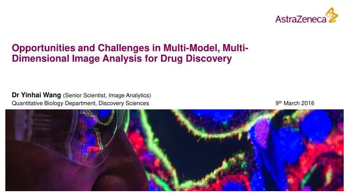
Opportunities and Challenges in Multi-Model, Multi- Dimensional Image Analysis for Drug Discovery Dr Yinhai Wang (Senior Scientist, Image Analytics) 9 th March 2016 Quantitative Biology Department, Discovery Sciences the Quantitative Biology
the Quantitative Biology Department (QuBi)
2
Mass Spectrometry Imaging (MSI)
3
Mass Spectrometry Imaging
What is Mass Spectrometry Imaging?
5
m 200 400 600 800
A single pixel in an MSI image is corresponding to a vector of numerical values. Fresh frozen sample an MSI image
What is Mass Spectrometry Imaging?
6
m 200 400 600 800
Why Mass Spectrometry Imaging?
7
- 1. MSI is able to detect and visualise the chemical composition.
- 2. MSI has ability to describe metabolite, drug concentration and biomarkers
simultaneously.
- 3. MSI is applicable to investigate drug’s efficacy and safety.
- 4. MSI is label-free.
m 200 400 600 800
Drug Biomarker
Relative Abundance Mass of Charge Ratio (m/z) – ion beam
Metabolite
MSI – Relationship Among Metabolite, Drug & Biomarker
8
*J. Cappell, University of Maastricht, unpublished work.
MSI + H&E
9
MSI images
Haematoxylin & Eosin (H&E) To look at the morphology, tissue structure
8 10 12 14 16 18 20 22 24 26 28 2 4 6 8 10 12 14 16 2 2.5 3 3.5 4 4.5 5 5.5 6 1.4 1.6 1.8 2 2.2 2.4 2.6 2.8 3 3.2
*J. Cappell, University of Maastricht, unpublished work.
H&E
10
H&E
11
- Y. Wang, et al. "Automated tumor analysis for molecular profiling in lung cancer." Oncotarget 6.29 (2015): 27938.
H&E + MSI (Metabolite)
12
- The spatial distribution of metabolite
- The correlation between tissue morphology with metabolite molecules
H&E + MSI (Drug)
13
Drug X
- The spatial distribution of drug X
- The relative concentration of drug X at different tissue locations
- The relationship between drug X and tissue morphology
H&E + MSI (Biomarker)
14
Biomarker A
- The location and spatial distribution of biomarker A
- The relative level of expression of biomarker A
- The relationship between biomarker A and tissue morphology
H&E + MSI (Metabolite, Drug, Biomarker)
15
Foreseeable problems:
- 1. Image Registration between MSI and H&E
- 2. Visualisation
- 3. Tissue image processing (e.g. machine
learning for the recognition of tumour *)
- 4. Multi-dimensional data analysis:
- Multiple MSI images
- MSI intensity + spatial locations
- H&E tissue structure
- Regional relationships
- Relationship among metabolite, drugs and
biomarkers)
- Machine learning for rapid matching
*Y. Wang, et al. "Assisted diagnosis of cervical intraepithelial neoplasia (CIN)." IEEE Journal of Selected Topics in Signal Processing, 3.1 (2009): 112-121.
Immunohistochemistry (IHC)
16
Fluorescent Multiplexing
17
MSI + H&E + Immunohistochemistry (IHC) + Fluorescent
18
IHC MSI
Fluorescent
H&E Multiple image modalities: m IHC + n H&E + p MSI + k Fluorescent
MSI for Dose Responses
19
Glutamine Glutamate Aspartate
Foreseeable problems: 1. Extra dimension of data 2. MSI Image registration (not from a same host) 3. The comparison of dose responses
MSI for Kinetics
20
Glutamine Glutamate Aspartate
Foreseeable problems: 1. Extra dimension of data 2. MSI Image registration (not from a same host) 3. The comparison of drug responses
- ver a period of time
The 3D Spheroid Model
21
85% of early clinical trials for novel drugs fail.
MSI for 3D Spheroid Models
22
Foreseeable problem: 3-dimensional image reconstruction Comparison of complex 3D image data
Liu, Xin, and Amanda B. Hummon. "Mass Spectrometry Imaging of Therapeutics from Animal Models to Three-Dimensional Cell Cultures."Analytical chemistry 87.19 (2015): 9508-9519.
3D slices
MSI Image Analysis
23
Multiple image modalities: m IHC + n H&E + p MSI + k Fluorescent Multiple image dimensions: 2D + colour channels + dose responses + kinetics + 3D
£1 million + 1TB + 6 months later
24
25
- MSI images are a stack of images.
– The matching between tissue morphology/structure (from H&E) and MSIs – The matching between IHC expression and MSIs – The matching between Fluorescent signals and MSIs
- We expect machine learning to play a key role in MSI image analysis, and be
aware of the following factors: – Expert input – Uncertainty – Unknown
What Machine Learning Could Do to Help?
26
Summary
27
1. At AstraZeneca, we are investigating novel imaging modalities. 2. We are generating a large amount of multi-modal image data, and in turn leads to a large amount of multi-dimensional numerical data. 3. We are pursuing better image and data analytical methods, machine learning approaches to
- Unleash the power of multiple image modalities (IHC + H&E + MSI +
Fluorescent)
- Unleash the power of multiple dimensional data (2D + colour channels +
dose responses + kinetics + 3D)
- To better understand biology and drugs’ mechanism of actions.
Acknowledgements
AstraZeneca - Quantitative Biology Department
- Claus Bendtsen
- Domingo Salazar
- Ian Barrett
- Lars Carlsson (Sweden)
- Johan Karlsson (Sweden)
AstraZeneca - High Content Biology Group
- Samantha Peel
- James Pilling
- Sinead Knight
AstraZeneca - Drug Safety & Metabolism
- Richard Goodwin
28
John Aston Carola Bibiane Schönlieb Ron Heeren Jo Cappell
Confidentiality Notice
This file is private and may contain confidential and proprietary information. If you have received this file in error, please notify us and remove it from your system and note that you must not copy, distribute or take any action in reliance on it. Any unauthorized use or disclosure of the contents of this file is not permitted and may be unlawful. AstraZeneca PLC, 2 Kingdom Street, London, W2 6BD, UK, T: +44(0)20 7604 8000, F: +44 (0)20 7604 8151, www.astrazeneca.com
29