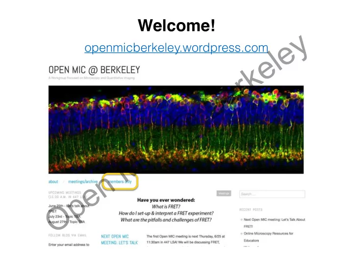
Open MIC @ Berkeley openmicberkeley.wordpress.com Open MIC @ - - PowerPoint PPT Presentation

open mic berkeley
SMART_READER_LITE
LIVE PREVIEW

Open MIC @ Berkeley openmicberkeley.wordpress.com Open MIC @ - - PowerPoint PPT Presentation

Welcome! Open MIC @ Berkeley openmicberkeley.wordpress.com Open MIC @ Berkeley Agenda Jen Lee: Introduction to FRET Marla Feller: Using FRET sensors to look at time resolved measurements Becky Lamason: Using FRET to determine if a


slide-1
SLIDE 1

Welcome!

  • penmicberkeley.wordpress.com

Open MIC @ Berkeley

slide-2
SLIDE 2

Agenda

  • Jen Lee: Introduction to FRET
  • Marla Feller: Using FRET sensors to look at time

resolved measurements

  • Becky Lamason: Using FRET to determine if a

bacterial protein manipulates cell-cell junctional tension

Open MIC @ Berkeley

slide-3
SLIDE 3

What is FRET?

Förster Resonance Energy Transfer 1946-1948, Theodor Förster Defined as: non-radiative, dipole-dipole resonance energy transfer

Open MIC @ Berkeley

slide-4
SLIDE 4

What is FRET?

Förster Resonance Energy Transfer Defined as: non-radiative, dipole-dipole resonance energy transfer i.e., no emission of a photon

http://nikon2.magnet.fsu.edu/articles/fluorescence/fret/fretintro.html

Open MIC @ Berkeley

slide-5
SLIDE 5

What is FRET?

Förster Resonance Energy Transfer Defined as: non-radiative, dipole-dipole resonance energy transfer

Ishikawa-Ankerhold, et. al., Molecules 2012, 17(4)

Open MIC @ Berkeley

slide-6
SLIDE 6

What is FRET?

Förster Resonance Energy Transfer Defined as: non-radiative, dipole-dipole resonance energy transfer

http://ascensionglossary.com/index.php/Law_of_Resonance

Open MIC @ Berkeley

slide-7
SLIDE 7

What is FRET?

Förster Resonance Energy Transfer Defined as: non-radiative, dipole-dipole resonance energy transfer

http://mlilm.iqfr.csic.es/materiales_laser_ing/index_ing.html

Open MIC @ Berkeley

slide-8
SLIDE 8

Why FRET?

  • “To detect interaction by spatial coincidence of

molecules” - Philippe Bastiaens (iBiology)

  • Molecular interactions within 10 nm range, better

resolution than traditional colocalization experiments

  • Allows for detection of dynamic events in vivo

(biosensors)

Open MIC @ Berkeley

slide-9
SLIDE 9

FRET Biosensors

http://zeiss-campus.magnet.fsu.edu/tutorials/spectralimaging/fretbiosensors/indexflash.html

Open MIC @ Berkeley

slide-10
SLIDE 10

Overlapping Spectra is Required for FRET, But Causes Bleedthrough

Open MIC @ Berkeley

slide-11
SLIDE 11

http://zeiss-campus.magnet.fsu.edu/articles/spectralimaging/spectralfret.html

Open MIC @ Berkeley

slide-12
SLIDE 12

FRET Efficiency

distance between donor & acceptor (nm) Förster Radius

k2 = orientation of transition dipoles J(l) = overlap integral of emission spectra n = refractive index of medium QD = quantum yield of donor

Open MIC @ Berkeley

slide-13
SLIDE 13

FRET Efficiency

Ishikawa-Ankerhold, et. al., Molecules 2012, 17(4)

Open MIC @ Berkeley

slide-14
SLIDE 14

Quantifying FRET = “Apparent Efficiency” (Eapp)

Eapp = E⍺

FRET efficiency geometric conformation distances & angles global parameter Donor-Acceptor Reaction [DA]/[Dtotal] How many molecules are in a complex? biologically relevant local parameter

Open MIC @ Berkeley

slide-15
SLIDE 15

How to Measure FRET

  • Sensitized Emission (Ratiometry)
  • Acceptor Photobleaching (Donor

Dequenching)

  • Fluorescence Lifetime Imaging Microscopy

(FLIM)

  • Spectral Imaging
  • Fluorescence Polarization Imaging (Anisotropy)

Open MIC @ Berkeley

slide-16
SLIDE 16

Measuring FRET: Sensitized Emission (Ratiometric Imaging)

  • Excite donor, then measure donor emission (DD) and

acceptor emission (DA)

  • Take the ratio of [DA/DD]
  • For more quantitative measurements, one can correct for

bleed through

DD DA Eapp= DA DD

Open MIC @ Berkeley

slide-17
SLIDE 17

Measuring FRET: Sensitized Emission (Ratiometric Imaging)

Wang, et. al., Molecular Imaging 12(2), 2013

Open MIC @ Berkeley

slide-18
SLIDE 18

Measuring FRET: Sensitized Emission (Ratiometric Imaging)

  • Pros:
  • Fast, good for live imaging
  • Easily implemented on standard scopes
  • Cons:
  • Very sensitive to noise
  • May require a lot of image processing (shade/flat-

field correction, bleedthrough correction, background subtraction, image alignment, photobleaching correction)

Open MIC @ Berkeley

slide-19
SLIDE 19

Measuring FRET: Acceptor Photobleaching (Donor Dequenching)

  • Excite donor, look at donor emission (DD). If FRET is
  • ccurring, then donor should be quenched.
  • Ask: what is the donor intensity in the absence of the

acceptor?

  • Bleach acceptor
  • Excite donor, then measure donor emission post-bleach

(DDpb).

  • Donor should unquench, resulting in higher intensity

from no FRET.

  • Eapp = 1 - (DD/DDpb)

Open MIC @ Berkeley

slide-20
SLIDE 20

Measuring FRET: Acceptor Photobleaching (Donor Dequenching)

Majoul, et. al., J Biotechnol. 2002;82(3).

Open MIC @ Berkeley

slide-21
SLIDE 21

Measuring FRET: Acceptor Photobleaching (Donor Dequenching)

  • Pros:
  • Easily implemented on standard scopes
  • No external calibration
  • Robust, reliable, and semi-quantitative
  • Cons:
  • Fixed samples or one time point only

Open MIC @ Berkeley

slide-22
SLIDE 22

Measuring FRET: Fluorescence Lifetime Imaging Microscopy (FLIM)

  • Every fluorophore has an exponential decay curve, a.k.a. a

fluorescence lifetime. When FRET occurs, the fluorescence lifetime of the donor decreases.

  • Process: excite donor, then measure donor lifetime (DD). If FRET,

then there will be faster donor decay.

http://nikon2.magnet.fsu.edu/articles/fluorescence/fret/fretintro.html

Open MIC @ Berkeley

slide-23
SLIDE 23

Measuring FRET: Fluorescence Lifetime Imaging Microscopy (FLIM)

  • Pros:
  • Direct measure of FRET efficiency
  • Independent of concentration
  • Acceptor doesn’t need to be imaged
  • Cons:
  • Specialized equipment required (we have one

instrument at the MIC!)

Open MIC @ Berkeley

slide-24
SLIDE 24

Measuring FRET: Spectral Imaging

  • Every fluorophore has a unique emission spectra.
  • Spectral imaging uses an array of highly sensitive

detectors to acquire intensities at specified wavelengths to plot out an emission spectra. Then, reference spectra are used to identify the fluorophore.

  • Similar approach to sensitized emission, but detection is

more specific.

Open MIC @ Berkeley

slide-25
SLIDE 25

Measuring FRET: Spectral Imaging

http://zeiss-campus.magnet.fsu.edu/articles/spectralimaging/spectralfret.html

Open MIC @ Berkeley

slide-26
SLIDE 26

Measuring FRET: Spectral Imaging

  • Pros:
  • Direct measure of FRET efficiency
  • Can correct for bleed through
  • Does not discard any signal (vs. sensitized emission)
  • Cons:
  • Specialized equipment required (we have many

instruments at the MIC!)

  • Requires reference spectra, controls

Open MIC @ Berkeley

slide-27
SLIDE 27

Measuring FRET: Polarization Anisotropy

https://www.microscopyu.com/articles/fluorescence/fret/fretintro.html

Open MIC @ Berkeley

slide-28
SLIDE 28

Measuring FRET: Polarization Anisotropy

  • Pros:
  • Independent of concentration
  • Relatively easy, inexpensive, and fast
  • Able to detect homo-FRET
  • Cons:
  • Not quantitative
  • Some common optical components destroy

polarization (e.g., high NA objectives)

Open MIC @ Berkeley

slide-29
SLIDE 29

Summary

  • FRET is a very powerful method of detecting

molecular interactions within 10 nm range.

  • No FRET approach is perfect. Goals, equipment,

and experimental system must all be considered.

  • FRET pairs should be chosen with care.

Open MIC @ Berkeley

slide-30
SLIDE 30

For more info…

  • iBiology FRET lecture by Philippe Bastiaens
  • Nikon, Zeiss, & Olympus education pages (all

written by Mike Davidson, Florida State University)

  • Cold Spring Harbor Quantitative Imaging Course

★ Links & slides will be available on the blog!

  • penmicberkeley.wordpress.com

Open MIC @ Berkeley