
SLIDE 1
New substrates for electron cryo-microscopy Lori Passmore 2014 - - PowerPoint PPT Presentation
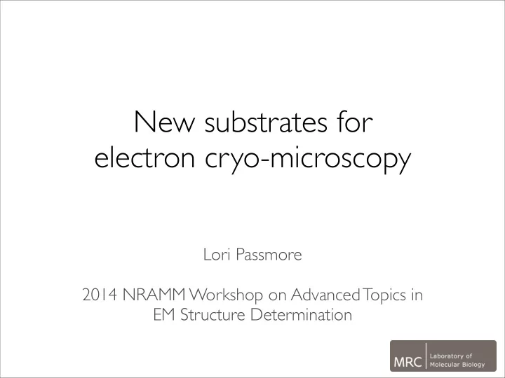
New substrates for electron cryo-microscopy Lori Passmore 2014 NRAMM Workshop on Advanced Topics in EM Structure Determination Tradi&onal substrates for cryo-EM electron microscope grid 80 m amorphous carbon membrane 1 m
metal grid bar amorphous carbon membrane ice embedded protein particles electron microscope grid 80 μm 1 μm
metal grid bar amorphous carbon membrane ice embedded protein particles electron microscope grid 80 μm 1 μm continuous
gold grid bar amorphous carbon membrane ice embedded protein particles electron microscope grid 80 μm 1 μm graphene
10 Å
Partial hydrogenation: Russo and Passmore (2014) Nature Methods Graphene oxide: Pantelic, Stahlberg et al (2010) JSB, (2011) JSB, (2011) Nano Lett Aromatic functionalisation: Pantelic et al (2014) Appl Phys Lett Amorphous carbon: Sader, Rosenthal et al (2013) JSB
95 90 85 80 75 70 65 Contact angle (degrees) 160 140 120 100 80 60 40 20 Hydrogen plasma exposure time (sec)
Russo & Passmore (2014) Nature Methods
0.143 0.5 1 10 6.1 5.1 5.2 5.0 3.6 2.7
1/Resolution (Å)
amorphous carbon: before motion correction after correction graphene: before motion correction after correction
b 80S
FSC
Amorphous carbon 0.18 Å/e–/Å2 0.47 Å/e–/A2 6 5 4 3 2 1 ) Å ( t n e m e c a l p s i d S M R 15 12 9 6 3 Fluence (e–/Å2) 600 300 Exposure time (ms) 900
Unsupported ice 0.14 Å/e–/Å2 0.50 Å/e–/Å2 15 12 9 6 3 900 600 300 Exposure time (ms) 15 900 Fluence (e–/Å2)
15 Graphene 0.092 Å/e–/Å2 0.41 Å/e–/Å2 15 12 9 6 3 900 600 300 Exposure time (ms) Fluence (e–/Å2)
EM, particularly as an alternative to thin amorphous carbon
reduces noise and radiation induced motion Russo & Passmore (2014) Nature Methods