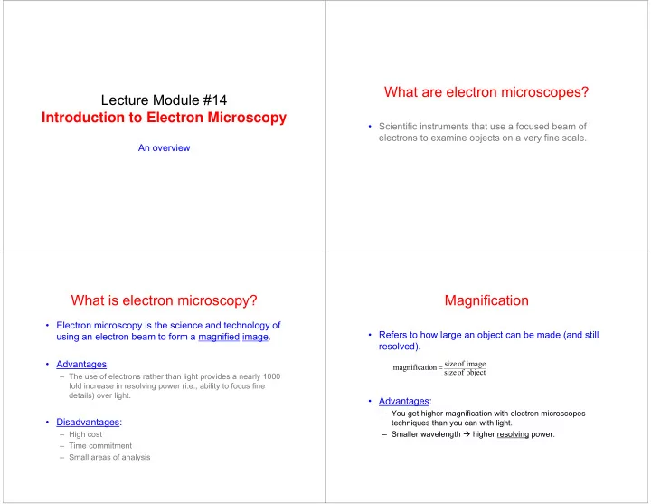

What are electron microscopes? Lecture Module #14 Introduction to Electron Microscopy • Scientific instruments that use a focused beam of electrons to examine objects on a very fine scale. An overview What is electron microscopy? Magnification • Electron microscopy is the science and technology of • Refers to how large an object can be made (and still using an electron beam to form a magnified image. resolved). • Advantages: size of image = magnificat ion size of object – The use of electrons rather than light provides a nearly 1000 fold increase in resolving power (i.e., ability to focus fine details) over light. • Advantages: – You get higher magnification with electron microscopes • Disadvantages: techniques than you can with light. – High cost – Smaller wavelength � higher resolving power. – Time commitment – Small areas of analysis
Resolution is: • The closest distance between two points that can clearly be resolved as separate entities through the microscope. Consider light as EM energy transmitted as a wave motion. We can consider Resolution is defined by: Intensity light as a series of r o ↓ ripples impinging upon an obstacle. Abbe relationship λ λ 0 . 61 0 . 61 d = 1 = = r Distance o α 2 sin n NA λ = wavelength of illuminant d 1 α = semi-angle n = index of refraction NA = numerical aperature B.D. Huey, MMAT322 Lecture Notes, University of Connecticut (2005) Important Terms Depth of Field • Depth of Field • A measure of how much of the object that we are – Height above and below the plane of focus that an image remains sharp. looking at remains in focus at the same time. – DOF is a function of magnification, α , and probe size • Advantages: BEAM Scan α – You get higher depth of field with (many) electron microscopy techniques than you do with light. Plane of focused image DOF – WHY? Region of image in focus
SEM SEM Other instruments we have or are getting TEM TEM • DualBeam Focused Ion Beam (DB-FIB) – Nano-machining platform • X-ray photoelectron spectrometer / Auger (XPS/Auger) EPMA EPMA • ??? Comparison of Selected Characteristics of Light What do we get out of electron and Electron Microscopes microscopes? FEATURE Light Microscope SEM TEM Surface morphology Surface morphology Sections (40-150 nm) Uses • Topography and sections (1-40 µ m) or small particles on thin membranes – Surface features of an object. “How it looks.” Source of Visible light High-speed electrons High-speed electrons Illumination • Morphology Best resolution 200 nm 3-6 nm 0.2 nm Magnification range 10-1,000 × 20-150,000 × 500-500,000 × – Size and shape of particles making up object. Depth of field 0.002-0.05 nm 0.003-1 mm 0.004-0.006 mm (NA=10 -3 ) (NA=1.5) • Composition Lens type Glass Electromagnetic Electromagnetic On eye by lens On CRT by scanning On phosphorescent Image ray- – Relative amount of elements and compounds making up the formation spot device screen by lens Information Phases Topography Crystal structure object. generated Reflectivity Composition Crystal orientation Crystal orientation Defects • Structure Composition Limiting Factor Wavelength of light Brighness, Lens quality – Crystallography. How atoms are arranged in the object signal/noise ratio, emission volume Adapted from: Scanning and Transmission Electron Microscopy, S. L. Flegler, J.W. Heckman Jr., K.L. Klomparens, Oxford University Press, New York, 1993.
History of electron microscopes All of this information is related to • Developed due to limitations of light optical properties. microscopes (LOMs) – LOM : ~1000x magnification; 0.2 µ m (200 nm) resolution properties • Transmission Electron Microscope (TEM) was developed first. – M. Knoll and E. Ruska, 1931 – Patterned “exactly” like a LOM. Uses electrons rather than structure processing light. • Scanning Electron Microscope (SEM) performance – 1942 Mechanical, Physical, Thermal, Optical, Electrical, etc… How do electron microscopes work? Goodhew, Humphreys & Beanland Electron Microscopy and Microanalysis, 3 rd Edition 70X Taylor & Francis, London, 2001 • Form a stream of electrons in the electron source (thermionic emission) and accelerate them towards the specimen using a positive electrical potential. 300X Electron microscopy • Use apertures and magnetic lenses to focus the provides significant stream into a thin focused monochromatic beam advantages over light optical microscopy in 1400X terms of resolution • Focus the beam onto the sample using another and depth of field magnetic lens. • Interactions occur inside the irradiated area of the 2800X sample. Collect results of interactions in a suitable detector and transform them into an image (or whatever you are interested in).
Electron Source The Electron Gun ( i.e. , electron gun) • The electron gun provides an intense beam of high Thermionic electron gun energy electrons. most widely used (~$70K-$150K) • There are two main types of gun. The thermionic Electrons are emitted gun, which is the most commonly used, and the field W Filament from a heated Bias emission gun. Wehnelt cap resistor filament and (negative potential) accelerated towards – • Electrons are generated and emitted from a filament Space charge an anode 10-1000 kV by thermionic emission (W or LaB 6 ) or from a sharp + Electron Beam tip by field emission (single crystal of W). Crossover Anode Plate (positive potential) • The electrons are accelerated through a potential Ground difference. Filament Operation of thermionic gun Made from a high T mp material with a low work function ( φ ) in Wehnelt order to emit as many electrons A biased grid with a potential that is as possible. The work function a few hundred volts different than • Apply a positive electrical potential to the anode is the energy needed by an the filament (cathode). This helps electron to overcome the barrier to accelerate the electrons and causes their paths to cross over. that prevents it from leaking out of the atom. φ W = 4.5 eV, φ LaB6 = • Heat the cathode (filament) until a stream of electrons is W Filament 3.0 eV. produced Bias Wehnelt cap – >2700 K for W resistor (negative potential) Crossover The effective source of illumination for the – • Apply a negative electric potential to the Wehnelt Space charge microscope. The size 10-1000 kV – electrons are repelled by the Wehnelt towards the optic axis is critical for high + Electron resolution Beam applications. Crossover • Electrons accumulate within the region between the filament tip Anode Plate and the Wehnelt. This is known as the space charge. (positive potential) Ground • Electrons near the hole exit the gun and move down the column Anode Positively charged metal plate to the target (in this case the sample) for imaging. at earth potential with a hole in it. It accelerates the electron beam to the high tension potential.
Other Types of Electron Sources ( i.e. , electron guns) The function of the electron gun is: To provide an intense beam of high Lanthanum hexaboride (LaB 6 ) energy electrons Higher brightness than W. More $$$ (~$500K) There are two main types of gun. The thermionic gun, which is the most commonly used, and the field emission gun. Brightness, B , is the beam Field emission source current density per unit solid Highest brightness. Even more $$$ (~$750K) angle. B = j c π β 2 ADVANTAGES j c = current density Provide higher current density → Higher brightness β = convergence angle http://www.matter.org.uk/tem/electron_gun/electron_sources.htm Interaction Volume Beam-Specimen Interaction • Represents the region penetrated by electrons • This is what makes electron microscopy possible. – Signals must escape the sample to be detected • Electrons strike the sample leading to a variety of reactions. Incident high kV beam of electrons Secondary e - Incident electron beam Characteristic Backscattered (SE) x-rays e - (BSE) Auger e - Visible light Bulk (SEM) e - - hole pairs Secondary electrons Absorbed e - Backscattered electrons Foil (TEM) Bremsstrahlung x-rays Interaction Volume (noise) X-rays Elastically Inelastically scattered e - scattered e - Direct (transmitted) beam • Use results of reactions to form image or generate other information.
Recommend
More recommend