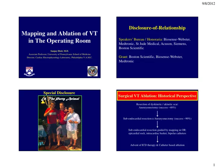

9/8/2012 Disclosure-of-Relationship Mapping and Ablation of VT Speakers’ Bureau / Honoraria: Biosense-Webster, in The Operating Room Medtronic, St Jude Medical, Acuson, Siemens, Boston Scientific Sanjay Dixit, M.D. Associate Professor, University of Pennsylvania School of Medicine Grant: Boston Scientific, Biosense-Webster, Director, Cardiac Electrophysiology Laboratory, Philadelphia V.A.M.C. Medtronic Special Disclosure Surgical VT Ablation: Historical Perspective Ed, The Party Animal Resection of dyskinetic / akinetic scar: Aneurysmectomy (success ~40%) Sub-endocardial resection ± Aneurysmectomy (success ~90%) Sub-endocardial resection guided by mapping in OR: epicardial sock, intracardiac basket, bipolar catheters Advent of ICD therapy & Catheter based ablation 1
9/8/2012 Surgical VT Ablation: Current Indications • Treatment of last resort for patients who have failed multiple Patient # Age (yrs) Sex LVEF (%) NICM ICD catheter ablation attempts. 1 65 M 15 Yes Yes 2 50 M 70 Yes Yes • PENN experience: over a 3 year period (2007 – 2009), 527 patients 3 54 M 58 Yes Yes underwent VT ablation – 295 patients (56%) had structural heart 4 51 F 25 Yes No disease (non-ischemic substrate in 144; 49%); 8 of these patients 5 51 M 30 Yes Yes (1.5%) needed surgical ablation. 6 74 M 30 Yes Yes 7 74 M 50 Yes Yes • All 8 patients has non ischemic cardiomyopathy: 6 with dilated cardiomyopathy and 2 with hypertrophic cardiomyopathy. 8 48 M 40 Yes Yes MRI identified septal scar in 3 patients; clinical VT was localized to thickened basal septum (>20 mm) in 2 of these subjects Intraseptal VT Substrate: MR Imaging Intra-Septal VT Substrate Mid Septal Scar Inferior Basal Scar Trans Thoracic Intra Cardiac RV LV Anterior Septal Scar Inferior Apical Scar 2
9/8/2012 ECG Features of VT originating from basal IVS Left bundle block with early precordial transition (V 1 – V 2 ) • Endocardial mapping was performed in all (LV in 8, RV in 4); epicardial mapping was performed in 6. • A total of 24 VTs (spontaneous / induced) were observed. • At conclusion of percutaneous ablation, in 4 patients no VT was Right bundle block inducible, in 3 patients clinical VT remained inducible and 1 with unusual precordial patient developed RV perforation requiring urgent surgery. transition • In all 4 patients that were non-inducible, ≥ 1 targeted VT recurred within a week of the last percutaneous ablation. Trans-Aortic View: Basal LV Cardiac Access for Surgical VT ablation AML Median Sternotomy provides best cardiac visualization: � RV epicardium LCC RCC � Superior IV Septum � Anterior LV wall NCC To visualize posterolateral LV wall, heart has to be physically lifted. Plane of Aortic Transaction Superior Anterior Posterior Partial sternotomy can allow visualization of RV, anterior and inferior LV surfaces. Inferior 3
9/8/2012 Surgical Ablation: Alternative Approaches VT Mapping in the OR • Trans-mitral approach : This offers best visualization of both papillary muscles, posterior LV endocardium and LV apex. • Finger mounted “roving” electrode: This was used for mapping critical components of the VT circuit in the early era of surgical ablation. • LV Apical approach : In patients with mechanical aortic and mitral valves. • Multipolar mesh / Basket catheter: High resolution so can provide comprehensive activation sequences; require special set-up • Aneurysmectomy site : Access to LV can be obtained through this for signal processing so cumbersome to use. location prior to aneurysm resection and closure. • Electro-anatomic mapping: Needs special set up – creation of • Accessing RV : Either via the tricuspid valve or the RV free wall. magnetic field, reference catheter (usually sutured to RV or LV epicardium). • Other approaches for epicardial access only : Partial sternotomy, window via epigastric incision, left anterior thoracotomy. Epicardial View Mapping in EP Lab prior to Surgical Ablation: PENN Approach • A priori detailed endocardial and when indicated, epicardial mapping performed in the EP laboratory. • Activation, entrainment and electro-anatomic mapping performed to identify critical components of VT circuit / site of origin vis-à- vis the underlying substrate. • These locations were targeted by conventional RF energy. • In the OR, these RF lesions were identifiable (especially endocardial) and served as targets for cryo-thermy applications. 4
9/8/2012 Trans-Aortic View Trans-Aortic View AML LCC RV & RVOT RCC NCC LCC RCC RFA Lesion (old) Superior Plane of Aortic Transaction Superior NCC RAA Anterior Posterior Anterior Posterior RFA Lesion (recent) Inferior Inferior Trans-Aortic Deployment of Cryo Probe Surgical VT Ablation: Lesion Creation Surgifrost™ Surgical Cryoablation System (Medtronic CryoCath LP, Quebec, Canada): The system uses Argon gas and can cool to -150 ° C. It utilizes flexible metal probe with adjustable insulation sheath. The standard duration of cryo-application is for a maximum of 3 minutes (cooling and thawing phases) and can create large lesions (~ 60 mm). Best results are achieved under cold cardioplegia which ensures adequate freezing. BASE LAT SEP APEX APEX RV LV BASE LAA 5
9/8/2012 Cryo Ablation: Final Result • Surgical approach utilized median sternotomy with cardio- pulmonary by pass under cold cardioplegia. • The epicardial and endocardial (via trans-aortic approach) surfaces were inspected and previously placed radiofrequency ablation lesions were identified. No additional mapping was performed in the OR. • Cryothermy ( Surgifrost, Medtronic CryoCath LP ) was applied to sites manifesting old lesions and / or scar identified in and around critical sites (temperature -150 ° C; total application time 3 minutes; anticipated lesion of 60 mm); additional cryo application on the opposing surface. Surgical VT Ablation: Acute End-points Lesion Creation in OR: Other Energy Sources • In cases where mapping / ablation are performed during ongoing VT, arrhythmia termination and non-inducibility should be the criteria. • Radiofrequency energy: Infrequently used for surgical VT ablation; may not be as effective in cooled hearts. • In cases where cold-cardioplegia is used during surgical ablation, heart requires rewarming in order to assess inducibility. VT induction can be influenced by deep sedation, anesthetic agents, cardiac filling, etc. • Laser energy (Nd-YAG, pulsed Argon): Have been used for surgical VT ablation in the past with excellent results; unclear why this modality is no longer used. • Other challenges: In patients undergoing concomitant valve or by-pass surgery and/ or experiencing de-compensation during surgical VT ablation, induction not advisable; lack of standard 12 lead ECG in OR may preclude • Microwave technology: Has also been shown to be effective for localization. lesion creation during surgical VT ablation. • Surrogates of substrate modification: non-capture, conduction block, etc. • Cryo-thermy: Remains the most common energy source for surgical VT ablation • Delayed (pre-discharge) assessment of VT inducibility. 6
9/8/2012 Surgical VT Ablation: Long-term Outcomes Patient # Time from NIPS pre- No. of AADs at No. of ICD surgery to discharge discharge shocks discharge (days) • Best long-term follow-up data: Patients who underwent surgical 1 11 Not performed 2 (Quinidine, Mexiletine) 3 VT ablation in the setting of healed myocardial infarcts: 1-year 2 5 Non-inducible 1 (Sotalol) 0 survival of 80-90% but by 5 th year 25% patients had died. 3 8 Not performed 0 0 4 Died N/A N/A N/A • PENN surgical VT Experience: All patients (n=8) had non- ischemic cardiomyopathy; 2 patients died during the hospitalization 5 7 Non-inducible 1 (Amiodarone) 0 following ablation; in remaining 6 patients over a mean follow-up 6 Died N/A N/A N/A 23±6 months, 4 patients were free of VT, 1 patient had single VT 7 6 Non-inducible 1 (Sotalol) 0 episode resulting in shock and 1 patient had 3 VT events in the first 3 months post-ablation but none after. 8 7 MMVT Inducible 1 (Mexiletine) 1 Surgical VT Ablation: Surgical VT Ablation: Conclusions Future Developments • Surgical ablation remains the treatment of last resort in • Ability to consistently induce and map VT in the OR patients experiencing VT that is refractory to conventional setting using the same tools as in the EP Lab: Hybrid OR. ablation. • Development of energy sources that can create effective • Ability to create large cryo lesions in the OR typically lesions without need for cold cardioplegia. under cold cardioplegia is the key to success of surgical VT ablation • Ability to map and ablate ventricular arrhythmias using a • Although mapping in the OR is ideal, a priori mapping and less invasive approach similar to what has been accomplished with surgical AF ablation. RF lesions in the EP lab can also guide surgical ablation 7
9/8/2012 Ed, we miss you at PENN………… Visually Identification of Epicardial Deployment of Cryo Probe Scar: • Infero-basal LV BASE • Lateral LV LAT SEP APEX APEX RV LV BASE LAA 8
Recommend
More recommend