little house on the prairie author wrote that her sister
play

Little House on the Prairie author wrote that her sister Mary was - PDF document

Background Encephalitis California Encephalitis Project (CEP) Case vignettes Highlights of agent-specific findings with focus on diagnostics (rather than Rx) Carol Glaser, DVM, MPVM, MD CEP experience and lessons


  1. • Background Encephalitis   California Encephalitis Project (CEP) • Case vignettes Highlights of agent-specific findings with focus on  diagnostics (rather than Rx) Carol Glaser, DVM, MPVM, MD  CEP experience and lessons learned, particularly as it relates to diagnostic testing Encephalitis and Special Investigations Section Present variety of cases-  Division of Communicable Disease Control  some relatively common where diagnostic problems California Department of Public Health arose and &  other rare, but important, causes Department of Pediatrics  Handout slightly different > lecture Division of Pediatric Infectious Diseases University of California, San Francisco • 
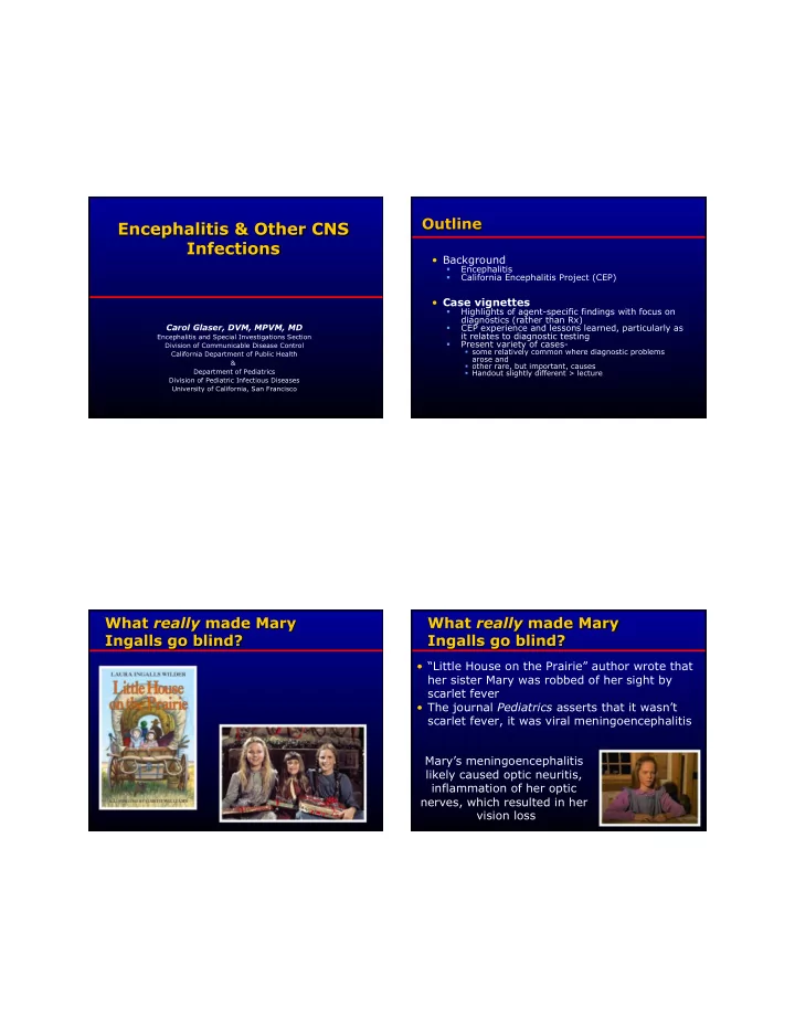
“Little House on the Prairie” author wrote that her sister Mary was robbed of her sight by scarlet fever • The journal Pediatrics asserts that it wasn’t scarlet fever, it was viral meningoencephalitis Mary’s meningoencephalitis likely caused optic neuritis, inflammation of her optic nerves, which resulted in her vision loss

  2. • Wide range of incidence rates depending on country, age-group etc., • 0.7-13.8/100,000 • Generally higher pediatric population > adults • Higher in tropical areas > “Western” countries • Comparable to ‘purulent meningitis’ Jmor F et al., Jour Virol 2008 Granerod J et al., Lancet Infect Dis 2010 Michael BD et al., Epilepsia, 2010 One of the most challenging syndromes for clinicians to diagnose and manage: • Severity of syndrome with high morbidity/ mortality • Vast number of infectious agents • Large number of non-infectious mimickers • Specific pathogen/underlying cause is identified < 50% of cases

  3. • Togavirus: EEE, VEE, WEE • Flavivirus: SLE, WN, JV, Dengue • Not a single disease entity • Bunyaviruses: LaCrosse, • Paramyxoviridae: mumps, measles • Often an uncommon presentation of a • Arenaviruses: LCM, Machupo, etc • Enteroviruses: Polio, coxsacki, etc common infection • Reoviruses: CTF • But sometimes a rare infection • Rhabdovirus: rabies • Filoviridae: Ebola, Marburg • Lots of misconceptions about diagnostic • Retroviridae: HIV • Herpes: HSV1/2,VZV,EBV,CMV,HHV6 testing • Adenovirus • Rickettsial • Bacterial • Fungal • Parasites • Prion • Non-infectious “ mimickers ”

  4. • Hospitalized w/ encephalopathy (depressed or ALOC > • 1998 – 2011 24 hours) • Viral and Rickettsial Disease Laboratory, AND State of CA • 1 or more of the following: • Funding from CDC Emerging Infections  fever (38 o C) Program  seizure(s)  focal neurological findings • Cases referred from MDs throughout CA  CSF pleocytosis  EEG findings c/w encephalitis Not population-based (e.g., large sampling   abnormal neuroimaging throughout CA)  Biased toward more severe and diagnostically • Exclusions: <6 months old or immunocompromised difficult cases • TN and NY had similar programs • CSF • Molecular, serologic, isolation • Acute serum • Multiple specimen types (CSF, sera, • Respiratory sample respiratory, brain if available) (NP/throat swab) • Core testing: Arboviruses (WNV, SLE, WEE) • Convalescent serum  Herpesviruses (HSV1, HSV2, VZ, EBV, HHV6)  (10-14 days > acute Enteroviruses  serum) Respiratory viruses (Flu A/B, Paraflu 1-3, adenovirus, HMPV)   Mycoplasma pneumoniae • Brain tissue if available • Expanded testing - exposures, clinical symptomatology, laboratory

  5. • Exposure history: • 10 year old, previously healthy,  Owns dog and cat white female Residence in rural area  No sick contacts  No recent travel   Admitted with 2 day history fever and upper respiratory illness, increasing • Admit labs/Neuroimaging lethargy and somnolence LP: WBC = 90 cells/mm 3 (75%L, 14%M),  Protein = 26 mg/ml, Glucose = 59 mg/ml  Admission exam - inattentive, drooling,  CT Scan: Left frontal lobe enhancement, and “ difficulty finding words ” mass effect

  6. CEP results • On HD#3 developed seizures • CSF PCR  HSV-1, HSV-2: Negative (HSV-1 PCR also negative • EEG: slowing L>R, sharp wave in left parietal outside hospital)  VZ: Negative  Mycoplasma : Negative • MRI: multifocal T2 prolongation with patchy  Enterovirus: Negative enhancement, most pronounced in left • Serology : temporal lobe  Arboviruses/ Mycoplasma /Chlamydia/ Adenovirus/EBV: Not significant • HD#4 LP repeated: • Respiratory PCR CSF WBC=113 WBC cells/mm 3 (83%L)   Influenza A/B, Adenovirus, Mycoplasma ,  Protein=107 mg/dl, Glucose=57 mg/dl Enterovirus: Negative 1. Rabies 2. Bacterial meningitis  CSF PCR HSV-1 repeated on 2 nd CSF: 3. Herpes simplex  Hospital lab: Positive HSV-1 encephalitis  VRDL: Positive HSV-1 4. Balamuthia mandrillis  Diagnosis: Herpes Simplex 5. Non-infectious, anti- Encephalitis (HSE) NMDAR 6. Enterovirus

  7. • CEP: 80 cases --~ 20% had initial PCR • HSV-1 considered to be leading negative (biased toward more difficult cases) cause of encephalitis • Of those with false negative 1 st CSF, CSFs • Acute necrotizing encephalitis were relatively bland: • PCR: considered sensitive and  Initial CSF lab values: specific  Median CSF WBC=17 WBCs/mm 3 (range: 0-330) - Tunkel AR et al., Clin Inf Dis, 2008  Median CSF Protein=34 mg/dL (range: 22-87) • False negative PCRs tend to occur early in course of illness (e.g., w/in 72 hours of onset) or CSF fluid relatively “ bland ’’  3/11 (27%) patients tested within 72 hours: negative-  repeat LP 5-11 day later positive - Weil AA et al, CID, 2002  8/33 (24%) negative first 3 days  5 cases repeat LP and 4 were positive - De Tiege X et al, CID, 2003  2/15 (13%) of HSE cases were initially negative HSV CSF PCR but results became positive in repeat CSF analyses - Elbers JM et al, Pediatrics, 2007

  8. • Exposure history • 10 year old male — Owns dog, cat and lizard — 3 days prior to admission with fever, right — Raccoons in yard sided weakness, and slurred speech — Plays football — No international travel, traveled to Central  On admission Valley  febrile  lethargy • Admit labs, neuroimaging  change in behavior — LP: WBC = 27 WBCs/mm 3 (100% Mo)  confusion — Protein = 23 mg/dL, Glucose = 54 mg/ml  somnolence — 1 st MRI: bilateral thalamic enhancement, L>R • Quickly deteriorated within first few • CSF PCR days of hospitalization  HSV1, HSV2: Negative  VZ: Negative — Intubated  Mycoplasma : Negative — Comatose  Enterovirus: Negative — Non-interactive Respiratory — Occasional — Enterovirus: Negative — Other viruses: Negative — Repeat MRI: area of enhancement • Serology: involvement with more extensive  Arboviruses (SLE, WEE, WNV), Mycoplasma , involvement of brainstem region Chlamydia, adenovirus, EBV: Not significant

  9. 1. Rabies • A. Rabies 2. Bacterial meningitis • B. Bacterial meningitis • C. Herpes simplex encephalitis 3. Herpes simplex • D. Balamuthia mandrillis encephalitis • E. Non-infectious, anti-NMDAR 4. Balamuthia mandrillis • F. Enterovirus 5. Non-infectious, anti- NMDAR 6. Enterovirus Most common infection identified : • Brain biopsy — “necrotizing encephalitis”, no • 26% of all confirmed etiologies: intranuclear cells, no organism 98 patients confirmed by positive PCR in CSF  • Median age = 14.0 years (mean=20.5 years) • Multiple studies done on biopsy • 63% pediatric, 37% adult — All negative — EXCEPT for weakly positive Entero • CSF: median WBC = 58 cells (0-2655), Protein=40 mg/dl (20-473), glucose=64 mg/dl (20-122) PCR — Prelim sequence data suggestive • Relatively short length of stay, median= 6 days EV 71 • Most with good outcomes, however 5+ fatalities (e.g. Enterovirus 71) Fowlkes AL et al., JID, 2008

  10. • 57 additional cases considered probable or Outbreak of Neurologic EV 71 possible –CSF EV PCR negative but positive in Disease: A Diagnostic Challenge respiratory and/or stool • CSF EV PCR good, but…not perfect -EV-PCR CSF ‘frequently negative’ -EV-PCR from resp and gi tract • Important to examine for enterovirus in have higher yields > EV-PCR CSF multiple specimens: Brain biopsy if possible  Throat/Nasopharyngeal  - Perez-Velez C, CID, 2007  Stool • Suspect we are missing EV cases due to unsuitable or incomplete specimens CSF Blood Throat Rectal other # specimens 16 3 6 8 9+ submitted # PCR 5 0 6 7 8 positive % 32% 0 100% 88% ~80% positive Perez-Velez C, CID, 2007

  11. • 12 year old previously healthy • Exposure history Bird at home Hispanic male   Motorcycling – dusty conditions  Presented with one week of progressively worsening headache, • Admission laboratory results vomiting, decreased appetite, poor  LP: WBC = 78 cells/mm 3 (78%L, 20%M), sleep, lethargy, and hallucinations Protein = 42 mg/ml, Glucose = 74 mg/ml  MRI suggestive of ADEM  CT concerning for brain tumor  Negative bacterial, fungal, & AFB stains & cultures  Intubated for increased ICP and ALOC Negative Coccidioides, Blasto, Histo, Cocci, &  Cysticercosis • Treatment • Symptoms returned  Decadron, Vancomycin, 2 weeks after stopping oral prednisone  Cefotaxime, Flagyl Headache, nausea, vomiting, and  lethargy • Repeat CT Worsening enhancement and edema  • Discharged one week later • Recurrent ADEM?  At baseline on 1 week oral Initial plan was for longer course of  prednisone taper with dx of ADEM steroids Changed after repeat MRI- concerning  for lymphoma

Download Presentation
Download Policy: The content available on the website is offered to you 'AS IS' for your personal information and use only. It cannot be commercialized, licensed, or distributed on other websites without prior consent from the author. To download a presentation, simply click this link. If you encounter any difficulties during the download process, it's possible that the publisher has removed the file from their server.

Recommend


More recommend