LETTER TO THE EDITOR T O THE E DITOR Here, we describe a case of HOD - - PDF document

letter to the editor
SMART_READER_LITE
LIVE PREVIEW

LETTER TO THE EDITOR T O THE E DITOR Here, we describe a case of HOD - - PDF document

LE JOURNAL CANADIEN DES SCIENCES NEUROLOGIQUES LETTER TO THE EDITOR T O THE E DITOR Here, we describe a case of HOD that presented acutely as a stroke mimic 14 months after a pontine cavernous malformation An Acute Clinical Presentation


slide-1
SLIDE 1

LE JOURNAL CANADIEN DES SCIENCES NEUROLOGIQUES

LETTER TO THE EDITOR

TO THE EDITOR An Acute Clinical Presentation Associated with Hypertrophic Olivary Degeneration Keywords: Hypertrophic olivary degeneration, Pontine caver- noma, Cavernous malformation, Guillain-Mollaret triangle, Stroke mimic Disruption of the Guillain-Mollaret triangle may result in hypertrophic olivary degeneration (HOD), resulting in palatal myoclonus, dysmetria, oscillopsia, dysarthria, and ataxia. Damage typically occurs within the dentate nucleus, dentatorubral tract, or rubro-olivary tract within the central tegmental tract,1 which is thought to result in deafferentation of the inferior olivary nucleus via trans-synaptic degeneration.2 Radiologically, a T2 hyperintense and hypertrophied inferior olivary nucleus is seen. Hypertrophy generally occurs 5-15 months after the pontine insult, while hyperintensity may occur as soon as 3 weeks post-insult.3 Here, we describe a case of HOD that presented acutely as a stroke mimic 14 months after a pontine cavernous malformation

  • resection. Hypertrophic olivary degeneration may result from

various pontine lesions,2 such as following the resection of a pontine cavernous malformation,4–6 which in one case resulted in a delayed, although not as acutely symptomatic, presentation of HOD.4 We present here the only known acute presentation of HOD following pontine cavernous malformation resection. We suggest that an acute presentation of HOD should be a rare but recognized complication of such resections to avoid unnecessary investigations, treatments, or procedures. A 58-year-old male was admitted to the neurology service 5 days after the acute onset of disequilibrium and gait dis-

  • turbance. He had undergone a left pontine cavernous mal-

formation resection via left temporal craniotomy 14 months

  • earlier. The surgery resulted in persistent dysarthria, right-sided

sensory impairment, and the requirement of a four-wheeled walker to ambulate. His deficits were stable until 5 days before his presentation, when he awoke and discovered that he was

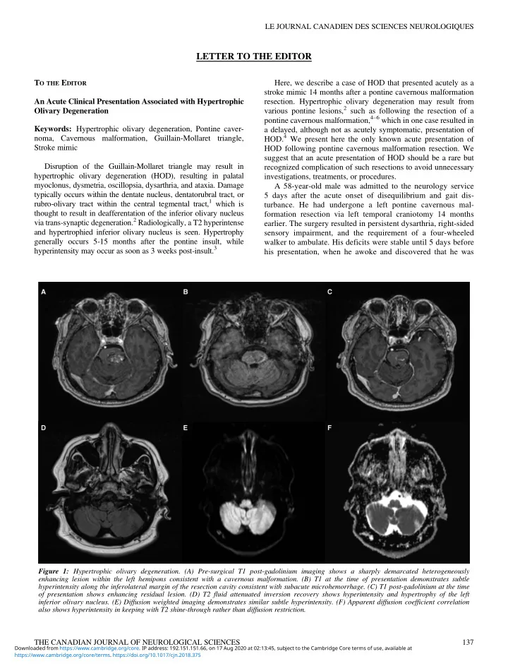
Figure 1: Hypertrophic olivary degeneration. (A) Pre-surgical T1 post-gadolinium imaging shows a sharply demarcated heterogeneously enhancing lesion within the left hemipons consistent with a cavernous malformation. (B) T1 at the time of presentation demonstrates subtle hyperintensity along the inferolateral margin of the resection cavity consistent with subacute microhemorrhage. (C) T1 post-gadolinium at the time

  • f presentation shows enhancing residual lesion. (D) T2 fluid attenuated inversion recovery shows hyperintensity and hypertrophy of the left

inferior olivary nucleus. (E) Diffusion weighted imaging demonstrates similar subtle hyperintensity. (F) Apparent diffusion coefficient correlation also shows hyperintensity in keeping with T2 shine-through rather than diffusion restriction. THE CANADIAN JOURNAL OF NEUROLOGICAL SCIENCES 137

https://www.cambridge.org/core/terms. https://doi.org/10.1017/cjn.2018.375 Downloaded from https://www.cambridge.org/core. IP address: 192.151.151.66, on 17 Aug 2020 at 02:13:45, subject to the Cambridge Core terms of use, available at

slide-2
SLIDE 2

unable to ambulate with his walker and had a constant sensation

  • f disequilibrium. He also noted a deterioration of his right-hand

coordination, exemplified by being unable to pour water from a jug without spilling. He denied visual changes, diplopia, new dysarthria, dysphagia, vertigo, tinnitus, hearing loss, weakness,

  • r new sensory impairment. There was no nausea, vomiting, or

headache. Neurologic examination was significant for a partial left abducens nerve palsy, bilateral gaze-evoked nystagmus, and upbeat nystagmus with upgaze. Saccades were hypometric to the left and hypermetric to the right. Facial sensation was decreased in V1, V2, and V3 of the left trigeminal nerve, but unchanged com- pared to post-cavernous malformation resection. There was no palatal myoclonus. Speech was dysarthric, but unchanged. There was a spastic catch in the left upper limb, but otherwise motor examination was normal. A rubral tremor was present in the right upper limb. There was 3+ hyperreflexia in the right brachioradialis, right biceps, and bilateral patellar reflexes. Plantar responses were

  • downgoing. Vibration sense was decreased in the right hemibody.

There was bilateral dysmetria on finger-nose and heel-shin testing that was more pronounced on the right. Dysdiadochokinesis was present in the right upper limb. Gait was assessed with assistance and was wide-based with irregularly spaced steps and marked ataxia

  • f the right leg. He was unable to perform tandem gait.

Owing to the acute onset of symptoms, computed tomography/ computed tomography angiography of the head and neck was performed to rule out a brainstem stroke. The findings showed the earlier resection bed, but no acute hemorrhage or vascular occlu-

  • sion. An enhanced MRI of the brain demonstrated a hypertrophied

T2 hyperintense (Figure 1D) left inferior olive with no diffusion restriction (Figures 1E and 1F). A small area of T1 hyperintensity near the prior resection site suggested subacute microhemorrhage (Figure 1B). The MRI findings were specific for hypertrophic degeneration

  • f the left inferior olive. Hypertrophy with degeneration is unique

to the inferior olivary nucleus. Therefore, non-enhancing, T1 isointense, T2 hyperintense hypertrophy of the lateral medulla in the region of the inferior olivary nucleus leaves essentially only the diagnosis of HOD. The finding of HOD on MRI explains our patient’s new right- sided dysmetria, ataxia, rubral tremor, and gait disturbance.7 Signs and symptoms typically manifest contralaterally to the lesion. Signs contralateral to the affected olive include dysmetria and limb dyscoordination. Other reported signs and symptoms include paresthesias, hypoesthesia, ataxia, dysarthria, diplopia, hemipar- esis, and lower limb spasticity.4–6 Many of these findings are likely a product of the inciting pontine lesion. Damage to the central tegmental tract and the encapsulated rubro-olivary tract results in ipsilateral HOD and contralateral symptoms.2 Palatal myoclonus, which was not present in our patient, is found in 7-33% of cases of HOD.1,2 In our patient, the previous cavernous malformation resection likely led to partial damage of the central tegmental tract. The acute

  • nset of symptoms in our patient may have been the result of

reaching a critical threshold of trans-synaptic degeneration. Alter- natively, a microhemorrhage may have damaged the remaining few axons in the rubro-olivary tract that had been maintaining a func- tionally complete circuit. Our patient was discharged from the acute neurology service for intensive inpatient rehabilitation. It is hoped that he will improve with time and physiotherapy, as has been reported in

  • ther cases.8

In summary, we present an acute clinical presentation associated with HOD from prior pontine cavernous malformation resection, with microhemorrhage as a possible explanation for the rapid onset

  • f symptoms. Hypertrophic olivary degeneration is a complication

that should be considered in patients presenting with acute symp- tomatology who have previously undergone brainstem cavernous malformation resection. Symptoms typically manifest con- tralaterally to the lesion, with variable rates of palatal myoclonus. Radiologic traits follow a well-defined time course, and are fairly specific should lateral medullary non-enhancing T2 hyperintense hypertrophy be seen at the level of the inferior olivary nucleus. DISCLOSURES Calvin Howard, Alexander Arnold, and Tyson Brust have nothing to disclose. STATEMENT OF AUTHORSHIP CH drafted the initial manuscript and assisted with figure

  • creation. AA provided editing and critical revisions to the

intellectual content as well as assisted with figure creation. TB provided editing and critical revisions to the intellectual content. Calvin Howard Undergraduate Medical Education Cumming School of Medicine University of Calgary, Calgary Alberta, Canada Alexander Arnold Department of Clinical Neurosciences Cumming School of Medicine University of Calgary, Calgary Alberta, Canada Tyson Brust Department of Clinical Neurosciences Cumming School of Medicine University of Calgary, Calgary Alberta, Canada Correspondence to: A. J. Arnold, Foothills Hospital, 12th Floor, C1245 1403-29th St N.W., Calgary, Alberta, Canada T2N 0B1. Email: arnoldaj@ucalgary.ca REFERENCES

  • 1. Gautier J C, Blackwood W. Enlargement of the inferior olivary

nucleus in association with lesions of the central tegmental tract or dentate nucleus. Brain. 1961;84:341-61.

  • 2. Jellinger K. Hypertrophy of the inferior olives. Zool Neurol.

1973;205:153-74.

  • 3. Kitajima M, Korogi Y, Shimomura O, Sakamoto Y, Hirai T,

Miyayama H, et al. Hypertrophic olivary degeneration: MR ima- ging and pathologic findings. Radiology. 1994;192(2):539-43. THE CANADIAN JOURNAL OF NEUROLOGICAL SCIENCES 138

https://www.cambridge.org/core/terms. https://doi.org/10.1017/cjn.2018.375 Downloaded from https://www.cambridge.org/core. IP address: 192.151.151.66, on 17 Aug 2020 at 02:13:45, subject to the Cambridge Core terms of use, available at

slide-3
SLIDE 3
  • 4. Hornyak M, Osborn AG, Couldwell WT. Hypertrophic olivary

degeneration after surgical removal

  • f

cavernous mal- formations of the brain stem: Report of four cases and review of the literature. Acta Neurochir (Wien). 2008;150 (2):149-56.

  • 5. Rosenblum JS, Nazari M, Al-Khalili Y, Potigailo V, Veznedaroglu E.

Unilateral symptomatic hypertrophic olivary degeneration sec-

  • ndary to midline brainstem cavernous angioma: A case report

and review of the literature. World Neurosurg [Internet]. 2018;110:294-300; Available from: https://doi.org/10.1016/j. wneu.2017.10.061.

  • 6. Yun JH, Ahn JS, Park JC, Kwon DH, Kwun BD, Kim CJ. Hyper-

trophic olivary degeneration following surgical resection or gamma knife radiosurgery of brainstem cavernous malformations: An 11-case series and a review of literature. Acta Neurochir (Wien). 2013;155(3):469-76.

  • 7. Lee S, Choi J, Son B. Rubral tremors associated with an inferior
  • livary lesion that developed after a brainstem hemorrhage. The
  • Nerve. 2015;1(1):50-2.
  • 8. Bouz P, Woods ROJ, Woods KRM. The pathophysiological basis for

hypertrophic

  • livary

degeneration (HOD) following brainstem insult. JSM Neurosurg Spine. 2013;1(1004):6-9. LE JOURNAL CANADIEN DES SCIENCES NEUROLOGIQUES Volume 46, No. 1 – January 2019 139

https://www.cambridge.org/core/terms. https://doi.org/10.1017/cjn.2018.375 Downloaded from https://www.cambridge.org/core. IP address: 192.151.151.66, on 17 Aug 2020 at 02:13:45, subject to the Cambridge Core terms of use, available at