
SLIDE 1
1
Pulmonary Function Testing – A Case Based Approach
Nitin Bhatt MD Karen Wood MD Pulmonary/Critical Care Medicine
Interpretation
- Is test acceptable and reproducible?
- Look at flow volume loop
- Examine FEV1/FVC ratio
- Look at FVC
- If obstruction – is there a post-bronchodilator
response
- Classify severity
- Look at lung volumes (specifically TLC)
- Examine DLCO
Interpretation
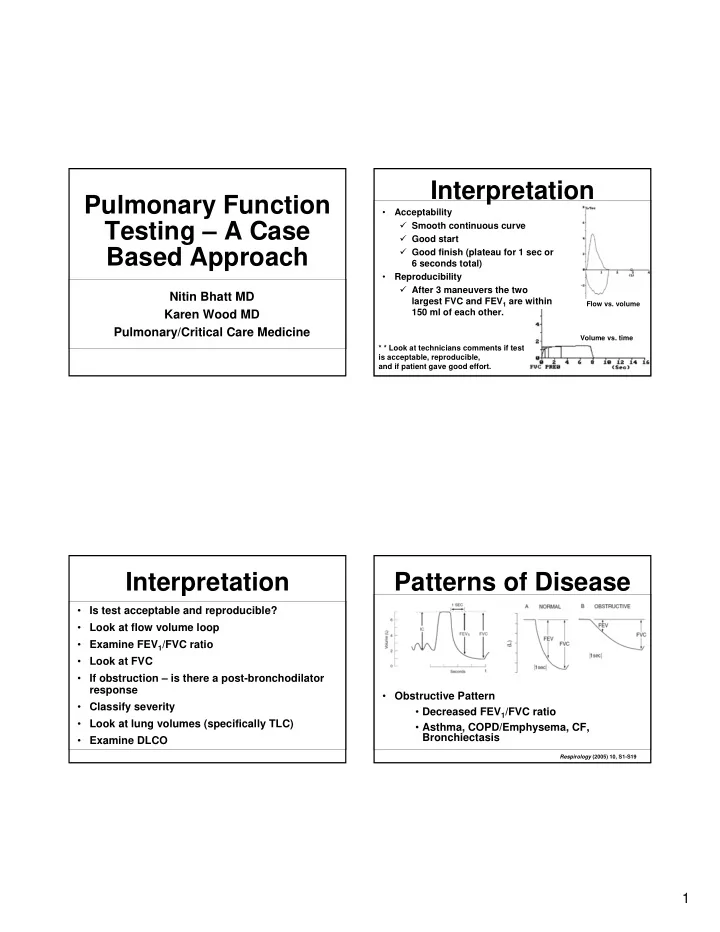
- Acceptability
Smooth continuous curve Good start Good finish (plateau for 1 sec or 6 seconds total)
- Reproducibility
After 3 maneuvers the two largest FVC and FEV1 are within 150 ml of each other.
Flow vs. volume Volume vs. time
* * Look at technicians comments if test is acceptable, reproducible, and if patient gave good effort.
Patterns of Disease
- Obstructive Pattern
- Decreased FEV1/FVC ratio
- Asthma, COPD/Emphysema, CF,
Bronchiectasis
Respirology (2005) 10, S1-S19