CASE STUDY CASE PRESENTATION: GD Pulmonary Hypertension Program - - PowerPoint PPT Presentation

case study
SMART_READER_LITE
LIVE PREVIEW

CASE STUDY CASE PRESENTATION: GD Pulmonary Hypertension Program - - PowerPoint PPT Presentation

Texas Childrens Hospital 4/21/2018 TCH128 PowerPoint System Process Round 6 Template Review CASE STUDY CASE PRESENTATION: GD Pulmonary Hypertension Program Pulmonary Hypertension Program DEPARTMENT NAME DEPARTMENT NAME 19 mo male


slide-1
SLIDE 1

Texas Children’s Hospital TCH128 PowerPoint System Process Round 6 Template Review 4/21/2018

DEPARTMENT NAME

Pulmonary Hypertension Program

CASE PRESENTATION: GD

DEPARTMENT NAME

Pulmonary Hypertension Program

CASE STUDY

DEPARTMENT NAME

Pulmonary Hypertension Program

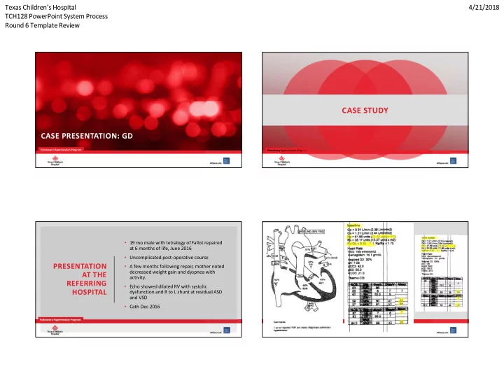
  • 19 mo male with tetralogy of Fallot repaired

at 6 months of life, June 2016

  • Uncomplicated post-operative course
  • A few months following repair, mother noted

decreased weight gain and dyspnea with activity.

  • Echo showed dilated RV with systolic

dysfunction and R to L shunt at residual ASD and VSD

  • Cath Dec 2016

PRESENTATION AT THE REFERRING HOSPITAL

DEPARTMENT NAME

Pulmonary Hypertension Program

slide-2
SLIDE 2

Texas Children’s Hospital TCH128 PowerPoint System Process Round 6 Template Review 4/21/2018

DEPARTMENT NAME

Pulmonary Hypertension Program

  • Remained intubated following cath, receiving inhaled nitric oxide and FiO2

1.0

  • Within 6 hours after cath, developed pulmonary edema, not responsive to

diuretics

  • Resolved with discontinuation of iNO
  • Strong concern for PVOD

DEPARTMENT NAME

Pulmonary Hypertension Program

  • CT chest showed changes of pulmonary

hypertension, not PVOD

  • No centrilobular nodules, no increased

septal thickening.

DEPARTMENT NAME

Pulmonary Hypertension Program

  • Lung biopsy: pathology was read locally and by consulting

pathologist as isolated pulmonary arterial hypertensive changes, normal venules

  • Not consistent with PVOD.

DEPARTMENT NAME

Pulmonary Hypertension Program

  • The child was stabilized and extubated
  • Discharged home on steroids, diuretics and oxygen as needed.
  • Worsening hypoxemia and work of breathing.
  • Inpatient admission for high flow nasal cannula support.
  • PH medications were not started because of poor clinical

tolerance.

  • Transferred to Texas Children’s Hospital for transplant

evaluation and pulmonary hypertension management (7/31/17).

slide-3
SLIDE 3

Texas Children’s Hospital TCH128 PowerPoint System Process Round 6 Template Review 4/21/2018

DEPARTMENT NAME

Pulmonary Hypertension Program

  • Maintained on high flow nasal cannula of 7 LPM at 25%
  • Comfortably tachypneic
  • He showed little response to increase in flow.
  • At night, supported with positive airway pressure for his

increased work of breathing (initial CPAP 6, changed to BPAP 12/6).

  • Goal saturations were 88-95%.
  • Steroids from the referring institution were weaned off.
  • Continued on bumetanide with additional doses of

diuretics as needed to maintain saturations and reduce work of breathing.

CLINICAL COURSE AT TEXAS CHILDREN’S

DEPARTMENT NAME

Pulmonary Hypertension Program

  • Review of CT chest: suspicious to medical team for PVOD; biopsy re-

evaluated

  • TCH radiology and pathology disagreement with PH team
  • PH medications not started
  • Lung transplant team consulted
  • Evaluation was completed and he was approved by the multidisciplinary

team for listing (8/10/17)

  • Insurance approval could not be obtained for transplant listing despite discussions,

letters and paperwork

DEPARTMENT NAME

Pulmonary Hypertension Program

  • The child acutely desaturated during a bowel movement,

requiring bagging and eventually, intubation for worsening acute respiratory failure (9 AM).

  • Developed pulmonary edema
  • Persistent hypoxemia
  • iNO, iloprost and oxygen given for rescue
  • Decreased cardiac output
  • Supported with epinephrine drip
  • Bradycardic event, no return of spontaneous circulation
  • 7 cycles of CPR with epinephrine, bicarbonate, calcium
  • Time of death: 10 AM

8/30/17

DEPARTMENT NAME

Pulmonary Hypertension Program

POST-MORTEM ANALYSES

slide-4
SLIDE 4

Texas Children’s Hospital TCH128 PowerPoint System Process Round 6 Template Review 4/21/2018

DEPARTMENT NAME

Pulmonary Hypertension Program

Lungs, features consistent with pulmonary veno-occlusive disease: 1. Interlobular septal vein, thickening with intimal sclerosis, variable occlusion, mild-moderate, patchy, bilateral. 2. Interlobular septal broadening and edema, venolymphatic ectasia, severe, diffuse, bilateral. 3. Intra-alveolar hemosiderosis, mild, patchy, bilateral. 4. No evidence of mutations in the EIF2AK4 gene by whole exome sequencing (Baylor Miraca Genetics). B. Secondary pulmonary arteriolar hypertensive changes, moderate-severe, diffuse, bilateral. C. No evidence of parenchymal fibrosis or infection. D. Congestive lymphadenopathy with sinus expansion, hilar and mediastinal, bilateral.

DEPARTMENT NAME

Pulmonary Hypertension Program

AUTOPSY

Occluded venule with partial recanalization, diagnostic for PVOD

DEPARTMENT NAME

Pulmonary Hypertension Program

  • Whole exome sequence negative for mutations (9/14/17)
  • Heterozygous for missense variants of uncertain clinical

significance on PAH mutation panel*

  • KCNA5
  • NOTCH3

* Texas Children’s PAH genetic panel: ACVRL1, BMPR2, CAV1, EIF2AK4, ENG, GDF2, KCNA5, KCNK3, NOTCH1, NOTCH3, SMAD4, SMAD8, TOPBP1

DEPARTMENT NAME

COMMENTS/QUESTIONS?

slide-5
SLIDE 5

Texas Children’s Hospital TCH128 PowerPoint System Process Round 6 Template Review 4/21/2018

DEPARTMENT NAME

Pulmonary Hypertension Program

FINAL DIAGNOSIS - PVOD

  • Rare form of pulmonary hypertension
  • 3-12% of those labeled idiopathic PAH
  • Estimated 1-2 cases per million
  • Preferential involvement of the

pulmonary venous system

  • Pathologic findings:
  • bliteration of small pulmonary veins by

intimal thickening

  • patchy capillary proliferation
  • Similar presentation to PAH but worse

prognosis.

  • Life threatening pulmonary edema

may occur following PH therapy

  • Heritable PVOD has been described
  • biallelic mutations of EIF2AK4
  • No effective treatments except for

lung transplantation