impact of disclosures who 2017 and ajcc 8 th edition
play

Impact of Disclosures WHO 2017 and AJCC 8 th edition Shareholder - PowerPoint PPT Presentation

5/26/2018 Impact of Disclosures WHO 2017 and AJCC 8 th edition Shareholder Five Prime Therapeutics and Adicet Bio on Gastropancreatic Neuroendocrine Neoplasms Consultant Celgene Grace E. Kim, MD These companies and its products will


  1. 5/26/2018 Impact of Disclosures WHO 2017 and AJCC 8 th edition Shareholder • Five Prime Therapeutics and Adicet Bio on Gastropancreatic Neuroendocrine Neoplasms Consultant • Celgene Grace E. Kim, MD These companies and its products will not be discussed in this presentation PanNEN WHO 2017 
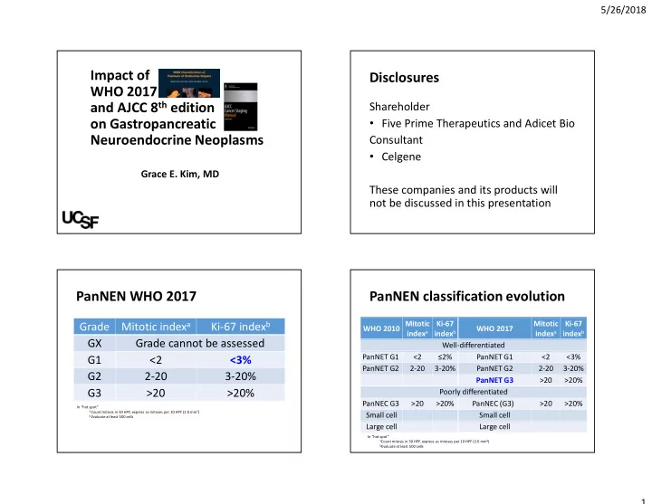
PanNEN classification evolution Mitotic index a Ki-67 index b WHO 2010 Mitotic Ki-67 Mitotic Ki-67 Grade WHO 2017 index a index b index a index b GX Grade cannot be assessed Well-differentiated PanNET G1 <2 ≤2% PanNET G1 <2 <3% G1 <2 <3% PanNET G2 2-20 3-20% PanNET G2 2-20 3-20% G2 2-20 3-20% PanNET G3 >20 >20% G3 >20 >20% Poorly differentiated PanNEC G3 >20 >20% PanNEC (G3) >20 >20% In “hot spot” a Count mitosis in 50 HPF, express as mitoses per 10 HPF (2.0 mm 2 ) Small cell Small cell b Evaluate at least 500 cells Large cell Large cell In “hot spot” a Count mitosis in 50 HPF, express as mitoses per 10 HPF (2.0 mm 2 ) b Evaluate at least 500 cells 1

  2. 5/26/2018 Disease specific survival of high grade panNENs Applicability All gastrointestinal and pancreatobiliary tumors • Primary and metastasis • Cytology, biopsy, and surgical resection Am J Surg Pathol. 2016; 40(9): 1192–1202. AJCC 8 th edition Neuroendocrine vs Carcinoma Neuroendocrine chapters Well differentiated Neuroendocrine • Ampulla of Vater and Duodenum neuroendocrine carcinoma (NEC) tumors (NET) • Appendix • WHO grade G1, G2 • High grade/ • Colon and Rectum and G3 poorly differentiated G3 • Small intestine • Stomach • Pancreas 2

  3. 5/26/2018 AJCC appendix chapters AJCC colon chapters Neuroendocrine Carcinoma Neuroendocrine Carcinoma VS VS pT1 ≤2 cm Invades submucosa Invades lamina propria or pT1 Invades submucosa submucosa and is ≤2 cm pT2 >2-4 cm Invades muscularis propria pT2 Invades muscularis propria or >4 cm or subserosal/ Through muscularis propria into Invades muscularis propria pT3 >2cm mesoappendix involvement subserosal/mesoappendix Invades muscularis propria Through muscularis propria into Perforates peritoneum or Invades visceral peritoneum/ pT3 into subserosa pericolorectal tissue pT4 directly invades adjacent serosa, directly invades adjacent organs/structures* organs/structures Invades visceral peritoneum Invades visceral peritoneum or pT4 (serosa) or other invades/adheres to adjacent *Excluding direct mural extension of adjacent bowel subserosa organs/structures organs/structures AJCC pancreas chapters Outline • Ki-67 index Neuroendocrine Exocrine VS pT1 <2 cm* ≤2 cm • G3 pancreatic neuroendocrine pT2 2 cm - 4 cm* >2cm - ≤4 cm neoplasms >4 cm* or invades pT3 >4 cm • Is this a mixed neoplasm? duodenum/bile duct Invades wall of large vessels Involves celiac axis, SMA, • Staging appendiceal neuroendocrine pT4 (celiac axis or SMA) or adjacent organ and/or common hepatic artery, (stomach, spleen, colon, adrenal gland) tumor regardless of size *Limited to the pancreas; extension of tumor into peripancreatic adipose tissue is NOT a basis of staging 3

  4. 5/26/2018 Outline To count • Ki-67 index • Check stromal cells are negative • G3 pancreatic neuroendocrine • Find labeling “hot spot” neoplasms • Evaluate at least 500 neoplastic cells • Is this a mixed neoplasm? • Report actual Ki67 index • Staging appendiceal neuroendocrine tumor Mod Pathol. 2016 Jan;29(1):93. Issues with Ki67 Figure from • Intensity can be variable • Due to antibody used and tissue section thickness • Weak nuclear stain? • If near categorical cutoffs • Count multiple “hot spots” and obtain average Mod Pathol. 2015;28(5):686-94. WHO classification of tumours of endocrine organs. World Health Organization; 2017. 4

  5. 5/26/2018 Surveyed colleagues WHO grade 2 Pathologist Circle + Slashes - % 28 383 6.8 Professor 1 28 383 6.8 Professor 2 32 415 7.7 Assistant Professor 3 39 499 7.8 Assistant Professor 4 30 419 7.2 Assistant Professor 5 36 425 8.4 GI Fellow 6 32 383 7.7 Which block? Look for • Architectural alteration • Confluent growth pattern with reduced tumor stroma and vasculature • Ischemic type tumor necrosis • Geographic, punctate, or single cell necrosis • Cytology changes • Increased nuclear size and atypia, nuclear membrane abnormalities, chromatin clumping • Mitotic activity Well-Differentiated Neuroendocrine Tumors with a Morphologically Apparent High-Grade Component: Clin Cancer Res. 2015 Feb 15;22(4):1011-7 5

  6. 5/26/2018 Outline Pancreatic NeuroEndocrine Neoplasms • Ki-67 index (PanNEN) • G3 pancreatic neuroendocrine neoplasms Pancreatic Pancreatic • Is this a mixed neoplasm? NeuroEndocrine Tumor NeuroEndocrine Carcinoma • Staging appendiceal neuroendocrine (PanNET) G3 (PanNEC) G3 tumor Both have mitotic >20/10 HPF and Ki67 >20% Clinical parameters at Treatment ends of NEN spectrum LG NET HG NEC NORDIC NEC study • Gastropancreatic NEN patients with Ki67 <55% are less Presentation Incidental finding Jaundice, weight loss, pain responsive to platinum agents Octreotide scintigraphy 18F-FDG-PET avid Imaging positive Serum ^ Neuroendocrine markers ^ Carcinoma markers Emerging data (chromogranin-A, gastrin, etc) (CEA, CA19.9, CA125) biomarkers • Both G3 panNENs respond to cytotoxic (platinum Clinical Protracted Rapid clinical deterioration and alkylating) agents (up to 10 years before recurrence) course Predictive and prognostic factors for treatment and survival in 305 patients with advanced gastrointestinal neuroendocrine carcinoma (WHO G3): the NORDIC NEC study. Ann Oncol. 2013 Jan;24(1):152-60. Pancreas 2017 Mar;46(3):296-301. 6

  7. 5/26/2018 Disease-specific survival (DSS) of stage-matched Does morphology distinguish? Clin Cancer Res. 2016 Feb 15;22(4):1011-7 WHO criteria of lung NEC 33 HG PanNEN cases Large cell Small cell Histologically ambiguous by Size /shape Large, round to polygonal <3x size of small resting lymphocyte, consensus in 20 cases (61%) round, oval, spindled Cytoplasm Moderate to abundant Scant, high nuclear/cytoplasmic ratio 1. All biopsies Cell border Ill-defined, prominent nuclear molding 2. Morphology of large cell Nuclear Vesicular Finely granular uniformly distributed “salt and pepper” or chromatin streaks chromatin NEC overlapped with WD NET Nucleoli Prominent, presence Absence to inconspicuous 3. One case had consensus facilitates separation from small cell NEC diagnosis of NEC was in fact Architecture Organoid nesting, palisading, rosettes, trabecula in both, also small WD NET / Necrosis cell NEC often has sheet-like growth with large areas of necrosis; Necrosis as abundant apoptotic cells or large prominent areas Am J Surg Pathol. 2016 Sep;40(9):1192-202. 7

  8. 5/26/2018 Morphologic clues for G3 Problematic NET • No clinical history • Appears well differentiated, but G3 detected by Ki67 • Limited sample • Co-existing or prior G1/G2 NET component • e.g. G1 panNET and G3 NET in liver • Not classic small cell NEC • Geographic necrosis but retained organoid pattern • Nested, trabecular, loosely cohesive but organized vascular network • No overt low grade component (G1/G2) and hyalinized intratumoral fibrosis NEC • Homogenous and lacks lower grade component • Co-existing conventional carcinoma • Expansile large irregular nests • Infiltrative, random large vessels, and desmoplastic type fibrosis Surveyed colleagues 8

  9. 5/26/2018 Pathologist Diagnosis Ki67 Me NEN (crush) 35% Professor 1 67% Professor 2 Favor LCNEC -> NEN (crush) >50% Assistant Professor 3 WD NET G3 46% Assistant Professor 4 NEN (crush) 45% Assistant Professor 5 WD NET G3 >20% GI Fellow 6 WD NET G3 -> NEN (crush) 54% Take home points 1.Try to count 2.Provide an actual % 3.Caveat if small or crushed sample Cytology PanNEC WD PanNET G1/G2 Large cell: Plasmacytoid WD PanNET G3 abundant cytoplasm, with round to ovoid Focally plasmacytoid large nuclei and nuclei, fine can have NEC features 87 HG NEC tubular GI tract vesicular nuclei or chromatin, (apoptosis/necrosis, prominent nucleoli smooth nuclear • 17/87 (20%) were neither small cell, large cell or mixed abundant cytoplasm, Small cell: membranes NEC large nuclei, prominent nuclear molding, nucleoli, molding) minimal cytoplasm, and hyperchromatic • “…the classic descriptions of small and large cell coarse, chromatin neuroendocrine carcinoma in the pulmonary Denovo G3 NEN when nonplasmacytoid system does not perfectly translate to the and has pleomorphic nuclei or abundant gastrointestinal tract and the pancreatobiliary system.” nucleoli requires mutational analysis Am J Surg Pathol. 2008 May;32(5):719-31. Cancer Cytopathol. 2018 Feb 16. [Epub ahead of print] Am J Surg Pathol. 2016 Sep;40(9):1192-202. Cancer Cytopathol. 2018 Jan;126(1):44-53. 9

  10. 5/26/2018 Case History 47 year old male with pancreatic NEN and bone and liver metastases status post neoadjuvant chemoradiation Synaptophysin 10

  11. 5/26/2018 Chromogranin Ki-67 Diagnosis? PanNEN genetic alterations • Pancreatic neuroendocrine tumor MEN1 DAXX/ATRX TP53 RB1 • Well-differentiated G3 NET WD NET 44% 43% 4% 0 • Pancreatic neuroendocrine carcinoma PD NEC 0 0 56% 72% • Large cell NEC Science. 2011 Mar 4;331(6021):1199-203. Am J Surg Pathol. 2012 Feb;36(2):173-84. 11

Download Presentation
Download Policy: The content available on the website is offered to you 'AS IS' for your personal information and use only. It cannot be commercialized, licensed, or distributed on other websites without prior consent from the author. To download a presentation, simply click this link. If you encounter any difficulties during the download process, it's possible that the publisher has removed the file from their server.

Recommend


More recommend