
SLIDE 1
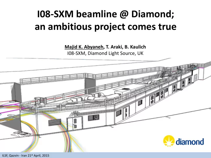
I08-SXM beamline @ Diamond; an ambitious project comes true
Majid K. Abyaneh, T. Araki, B. Kaulich I08-SXM, Diamond Light Source, UK
ILSF, Qazvin - Iran 21st April, 2015
I08-SXM beamline @ Diamond; an ambitious project comes true Majid - - PowerPoint PPT Presentation
I08-SXM beamline @ Diamond; an ambitious project comes true Majid K. Abyaneh, T. Araki, B. Kaulich I08-SXM, Diamond Light Source, UK ILSF, Qazvin - Iran 21 st April, 2015 Villages Phase I Phase II Phase III Macromolecular I02 I04-1 VMXi
Majid K. Abyaneh, T. Araki, B. Kaulich I08-SXM, Diamond Light Source, UK
ILSF, Qazvin - Iran 21st April, 2015
Villages Phase I Phase II Phase III Macromolecular Crystallography I02 I03 I04 I04-1 I24 VMXi VMXm I23 Spectroscopy I18 (I18) I20 B18 I08 (I08) I14 (I14) I21 Soft Condensed Matter B23 I22 B22(B22) B21 Materials I16 I13 (I13) I19 B16 Engineering & Environment I15 I12 (I12) I11 I15-1 Surfaces & Interfaces I06 (I06) I07 I10 I09 B07 I05 Imaging B24
ILSF, Qazvin - Iran 21st April, 2015
B24 B16 I12: 53-150 keV; < 2-3 mm
ILSF, Qazvin - Iran 21st April, 2015
Bridge-DH
ILSF, Qazvin - Iran 21st April, 2015
HU53
ILSF, Qazvin - Iran 21st April, 2015
Control Rooms 1&2/ Specimen Prep Area (±0.5 oC; <40 dB; Office environment) Experiments Cabin (±0.1 oC; <40 dB; Lab environment) Optics Cabin II (±0.5 oC; <45 dB; Optics Cabin I (±0.5 oC; <45 dB) Optics Hutch (±0.5 oC; <50 dB) Airlock Vestibule
Support Lab
ILSF, Qazvin - Iran 21st April, 2015
ILSF, Qazvin - Iran 21st April, 2015
Ni coating stripe Au coating stripe Measurement RMS (Å) PV (Å) RMS (Å) PV (Å) 1 1.46 53 0.98 33 2 0.86 39 0.97 33 3 0.83 40 0.95 36 4 1.05 58 0.88 36 5 0.91 42 0.93 33 Average 1.02 46 0.94 34
Figure 1: Examples of the surface roughness figures (the first two measurements) of the Au stripe. Table 1: The measurement result.
(Removed term: the 2nd order of Zernike polynomial)
Figure 2: Examples of the surface roughness figures (the first two measurements) of the Ni stripe.
Au Ni
ILSF, Qazvin - Iran 21st April, 2015 Sq=Root Mean Square; Sy=Height of Peak to Valley
Manufacturer: Gooch & Housego
ILSF, Qazvin - Iran 21st April, 2015
10 mm exit slit; cff=4
ILSF, Qazvin - Iran 21st April, 2015
1- Pre-PGM version :water-cooled 2- Post-PGM version :un-cooled
ILSF, Qazvin - Iran 21st April, 2015
in
Static, 4m long granite End station on floating granite Exit slit/ Secondary source Attenuator stage Diagnostics unit
ILSF, Qazvin - Iran 21st April, 2015
Transmission EMCCD detector system LN2 cryogenic cooling and transfer system X-ray fluorescence detector In-situ visible light microscope Control system for vacuum and cryo transfer
ILSF, Qazvin - Iran 21st April, 2015
Zone Plate, OSA, Central Stop,...
CCD, SDD, Electron analyser
Sample stage
OSA FZP Feedthrough and cabling
ILSF, Qazvin - Iran 21st April, 2015
Vacuum window ZP stage with scanner XRF detector OSA Laser interferometer Transmission detector system Specimen coarse and fine stage Cryo stage Cold trap
ILSF, Qazvin - Iran 21st April, 2015
ray diffraction, XANES IRD Quad segment photo diode Transmission imaging with simultaneous acquisition
Bruker X-Flash 6|100 XRF detector (with a single 100mm2 SDD detector and XIA MCA) Elemental mapping
Conventional set-up
Fibre-optic coupled
ILSF, Qazvin - Iran 21st April, 2015
ILSF, Qazvin - Iran 21st April, 2015
Specimen environment Description Standard dry specimen Kinematic mount with repositioning precision of at least 5 um or less than 10%
Cryogenic specimen environment LN2 cooled specimen (< -160 oC) with temperature stability < 0.1 oC including transfer system Electrochemical reaction cell (in design phase) Closed chemical reaction cell with electrical biasing, in- and outlet of reactants (liquid and gases) Chemical reactors for new biomaterials (in design phase) Closed chemical microfluidic reaction cell with pH and temperature control Micro heater (under commissioning) Microheater on Si3N4 membrane with temperature sensor (200 oC on day-1; max 500 oC) Magnetic field Specimen with remanent magnetic field, no external magnetic field applicable
ILSF, Qazvin - Iran 21st April, 2015
Plunge freezing the grids by Leica EM GP
Cryo-stage Not ordered yet
Cryo Microtom
ILSF, Qazvin - Iran 21st April, 2015
Mounting and loading samples under LN2
ILSF, Qazvin - Iran 21st April, 2015
ILSF, Qazvin - Iran 21st April, 2015
1st order diffraction cone
15x15 mm 8x6 mm
ILSF, Qazvin - Iran 21st April, 2015
I08-SXM: Status update - Imaging Test pattern from zoneplates.com; 720 eV photon energy, 400px x 400px, 1ms dwell/s; Smallest features in the Siemens star are 50 nm 2 mm
ILSF, Qazvin - Iran 21st April, 2015
2 mm
ILSF, Qazvin - Iran 21st April, 2015
I08-SXM: Status update - Imaging nucleous perinuclear region cytosol membrane
neuropil Rat cortical neuron cell in brain tissue; 34x36 um, 845 eV Courtesy of C. Poitry-Yamate @ CIBM, EPFL Lausanne, CH 720 eV
ILSF, Qazvin - Iran 21st April, 2015
N
Transmission Horizontal Vertical Courtesy of C. Poitry-Yamate @ CIBM, EPFL Lausanne, CH
C Fe O
2 mm 2 mm
C Fe N cryo-XRF map; Ferihydrate co-prep Geobacter sulfurreducens T < 160 C Courtesy of Prof. J. Lloyd; University of Manchester
ILSF, Qazvin - Iran 21st April, 2015
NEXAFS spectromicroscopy on Fe nano
Keele University.
Fe
To explore biomineralised iron deposits in organic materials
ILSF, Qazvin - Iran 21st April, 2015
Transmission imaging and XRF mapping of particles from a hydrothermal vent orifice in the deep sea; samples provided by Rachel Mills, Southampton University
http://www.pmel.noaa.gov/eoi/ XRF mapping of particles from deep-sea vents
(D. Wang, RSC, 2013) (D. Wang, Crys Growth Des, 2011) Extracted spectra from 6 images around Sn-edge
STXM images with 30nm resolution @510 eV
480 eV 495 eV 510 eV
200 nm
A B
1 um
ILSF, Qazvin - Iran 21st April, 2015
CNTs on the ITO surface
Ptychographic reconstruction
images of a mouse fibroblast cell doped with cobaltferrite ENPs (pilot experiment performed at TwinMic, Elettra) Collaboration with Sheffield University and University College London
ILSF, Qazvin - Iran 21st April, 2015
Visit us at: http://www.diamond.ac.uk/Beamlines/Spectroscopy/I08.html Follow us on: https://www.facebook.com/DiamondLightSource https://twitter.com/DiamondLightSou/ I08-SXM local Contacts:
burkhard.kaulich@diamond.ac.uk
majid.abyaneh@diamond.ac.uk
ILSF, Qazvin - Iran 21st April, 2015