# of true positives true positive rate = # of known positives - - PowerPoint PPT Presentation

of true positives true positive rate of known positives
SMART_READER_LITE
LIVE PREVIEW

# of true positives true positive rate = # of known positives - - PowerPoint PPT Presentation

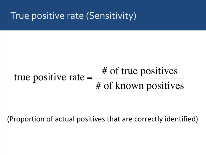
True positive rate (Sensitivity) # of true positives true positive rate = # of known positives (Proportion of actual positives that are correctly identified) True negative rate (Specificity) # of true negatives true negative rate = # of known


slide-1
SLIDE 1

True positive rate (Sensitivity)

true positive rate = # of true positives # of known positives

(Proportion of actual positives that are correctly identified)

slide-2
SLIDE 2

True negative rate (Specificity)

true negative rate = # of true negatives # of known negatives

(Proportion of actual negatives that are correctly identified)

slide-3
SLIDE 3

False positive rate (1 – Specificity)

false positive rate = # of false positives # of known negatives

(Proportion of actual negatives that are incorrectly identified)

slide-4
SLIDE 4

Sensitivity and specificity depend on a chosen cutoff

cutoff malignant benign false positives false negatives

slide-5
SLIDE 5

Sensitivity and specificity depend on a chosen cutoff

cutoff malignant benign false negatives false positives

slide-6
SLIDE 6

Do Part 1 of the worksheet now

slide-7
SLIDE 7

We usually plot the true pos. rate vs. the false

  • pos. rate for all possible cutoffs

ROC curve Receiver Operating Characteristic curve

slide-8
SLIDE 8

Image from: http://en.wikipedia.org/wiki/Receiver_operating_characteristic

slide-9
SLIDE 9

The area under the curve tells us how good a model’s predictions are

worst case good perfect

slide-10
SLIDE 10

Let’s look at the performance of several different models for the biopsy data set

slide-11
SLIDE 11

Predictor M1 clump_thickness normal_nucleoli marg_adhesion bare_nuclei uniform_cell_shape bland_chromatin

slide-12
SLIDE 12

Predictor M1 M2 clump_thickness normal_nucleoli marg_adhesion bare_nuclei uniform_cell_shape bland_chromatin

slide-13
SLIDE 13

Predictor M1 M2 M3 clump_thickness normal_nucleoli marg_adhesion bare_nuclei uniform_cell_shape bland_chromatin

slide-14
SLIDE 14

Predictor M1 M2 M3 M4 clump_thickness normal_nucleoli marg_adhesion bare_nuclei uniform_cell_shape bland_chromatin

slide-15
SLIDE 15

Predictor M1 M2 M3 M4 M5 clump_thickness normal_nucleoli marg_adhesion bare_nuclei uniform_cell_shape bland_chromatin

slide-16
SLIDE 16

Model Area Under Curve (AUC) M1 0.909 M2 0.968 M3 0.985 M4 0.995 M5 0.996

slide-17
SLIDE 17

Things usually look much worse in real life

Keller, Mis, Jia, Wilke. Genome Biol. Evol. 4:80-88, 2012

Best AUC (solid line): 0.70

slide-18
SLIDE 18

Do Part 2 of the worksheet now