
SLIDE 1
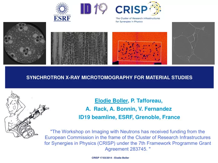
SYNCHROTRON X-RAY MICROTOMOGRAPHY FOR MATERIAL STUDIES
Elodie Boller, P. Tafforeau,
- A. Rack, A. Bonnin, V. Fernandez
ID19 beamline, ESRF, Grenoble, France
CRISP 17/03/2014 - Elodie Boller
"The Workshop on Imaging with Neutrons has received funding from the European Commission in the frame of the Cluster of Research Infrastructures for Synergies in Physics (CRISP) under the 7th Framework Programme Grant Agreement 283745. "